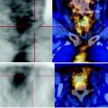
Fig. 17.1
The PET-CT examination demonstrates modest thickening tissue with high carbohydrate metabolism in correspondence of the rectal anastomosis adjacent to the metal clips, in contiguity with the posterior wall of the bladder
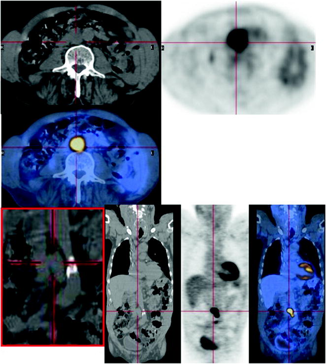
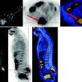
Fig. 17.2


The PET-CT scan shows a conglomerate of lymph nodes measuring 22 x 25 mm in the axial plane and characterized by high metabolism. This mass extends from the intercavo-aortic region, through a plane passing over the renal arteries, reaching the aorto-iliac bifurcation
Stay updated, free articles. Join our Telegram channel

Full access? Get Clinical Tree


