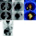
Fig. 31.1
Axial PET-CT images show some enlarged lymph nodes increased in size in the presacral left region
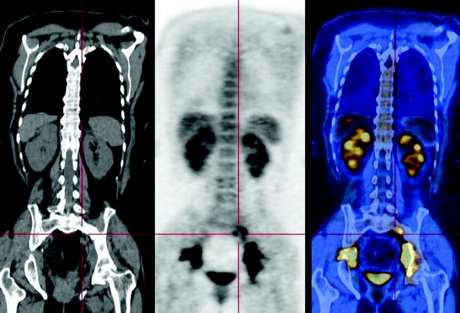
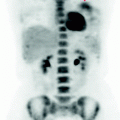
Fig. 31.2
 Laryngeal Squamous Carcinoma: Staging
 Radio-Treated Cancer of the Posterior Hemi-Circumference of the Anal Canal: Post-Actinic Fibrosis
 Bone-Destroying Metastases in Thyroid Undifferentiated Carcinoma
 Lymphocytic Interstitial Pneumonia in Patient with History of Breast Cancer
 Metastatic Breast Carcinoma: Restaging After Neoadjuvant Chemotherapy
 Bone Metastases from Breast Cancer: Progression of Disease and Subsequent Response to Radiotherapy


The coronal reconstructions confirm the presence of adenopathy in the presacral and better demonstrate the involvement of the obturatory stations on both sides, also characterized by high metabolism (see also Fig. 31.3)
Related posts:
Laryngeal Squamous Carcinoma: Staging
Radio-Treated Cancer of the Posterior Hemi-Circumference of the Anal Canal: Post-Actinic Fibrosis
Bone-Destroying Metastases in Thyroid Undifferentiated Carcinoma
Lymphocytic Interstitial Pneumonia in Patient with History of Breast Cancer
Metastatic Breast Carcinoma: Restaging After Neoadjuvant Chemotherapy
Bone Metastases from Breast Cancer: Progression of Disease and Subsequent Response to Radiotherapy
Stay updated, free articles. Join our Telegram channel
Full access? Get Clinical Tree
