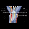
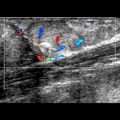
Swollen Nerve
ESSENTIAL INFORMATION
Key Differential Diagnosis Issues
Helpful Clues for Common Diagnoses
 Tumor of peripheral nerve
 Hypoechoic, fusiform-shaped tumor of nerve
 Thickening of entering or exiting nerve
 Anechoic areas
 Posterior acoustic enhancement
 Moderately hyperemic on Doppler
 ± hyperechoic areas
 Malignant peripheral nerve sheath tumor is more likely if
 FDG PET has quite high (~ 80-90%) sensitivity and specificity for diagnosing malignant peripheral nerve sheath tumor associated with neurofibromatosis
Helpful Clues for Less Common Diagnoses
 Chronic inflammatory demyelinating polyneuropathy (CIDP) and multifocal mononeuropathy (MMN) are acquired immune-mediated peripheral neuropathies


