Synovial Cyst


Clinical Presentation


The patient is a 58-year-old man with 4- to 6-week history of low back pain, bilateral buttock pain, right lower extremity pain, numbness of both feet, and difficulty walking. He has weakness in both hip extensors, both hip abductors, and bilateral weakness of the foot, right greater than left. Clinical findings are consistent with multilevel radiculopathy and neurogenic claudication.


Imaging Presentation


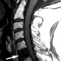
Magnetic resonance (MR) imaging of the lumbar spine reveals a large, lobulated synovial cyst arising from the anteromedial aspect of the right L4-5 facet joint. The cyst encroaches upon the medial aspect of the right L4-5 neural foramen and causes marked compression and displacement of the thecal sac from right to left ( Figs. 75-1 to 75-6 ) .


Figure 75-1


Large Synovial Cyst, L4 Level.

Right parasagittal T2-weighted MRI. A large fluid-filled synovial cyst ( long arrow ) is positioned above the L4-5 intervertebral disc level in the spinal canal posterior to the L4 vertebral body. Noted is a Schmorl’s node ( short arrow ) in L3 vertebral body inferiorly.


Figure 75-2


Large Synovial Cyst, L4 Level.

Same patient as in Figure 75-1 . Corresponding right parasagittal T1-weighted MRI. The synovial cyst ( long arrow ) is nearly isointense relative to intrathecal CSF. Noted is Schmorl’s node ( short arrow ) in L3 vertebral body inferiorly.


Figure 75-3


Large Synovial Cyst, L4 Level.

Same patient as in Figures 75-1 and 75-2 . Right parasagittal contrast-enhanced fat-saturated T1-weighted MRI. The synovial cyst contains fluid that is isointense relative to CSF. The margins of the cyst enhances intensely ( long arrow ). The L3 Schmorl’s node ( short arrow ) enhances intensely.


Figure 75-4


Large Synovial Cyst, L4 Level.

Axial contrast-enhanced fat-saturated T1-weighted MRI obtained just above L4-5 intervertebral disc level in same patient as in Figures 75-1 to 75-3 . The cyst margins ( arrows ) enhance with contrast, but fluid in the cyst does not enhance. The cyst extends into the medial aspect of the right L4-5 neural foramen ( short arrow ). A portion of the cyst is located in the spinal canal ( long arrow ) where it displaces the thecal sac to the left.


Figure 75-5


Large Synovial Cyst, L4 Level.

Axial contrast-enhanced T2-weighted MRI obtained just above L4-5 intervertebral disc level in same patient as in Figure 75-4 . The cyst ( arrows ) is lobulated and contains T2 hyperintense fluid. The infrapedicular portion of the cyst ( short arrow ) extends into the medial aspect of the right L4-5 neural foramen where it encroaches upon the right L4 nerve root ( L4 ). The portion of the cyst in the spinal canal ( long allow ) compresses and displaces the thecal sac ( T ) toward the left.


Figure 75-6


Large Synovial Cyst, L4 Level.

Axial unenhanced T1-weighted image A and axial contrast-enhanced fat-saturated T1-weighted MR image B , obtained at the L4-5 facet level, in same patient as in Figures 75-1 to 75-5 . The synovial cyst ( short arrow in A and B ) obscures the fat in the medial aspect of the right L4-5 neural foramen. The facets ( long arrows in image A ) are hypertrophic secondary to osteoarthritis. The posterior facets and facet capsules ( long arrows in B ) enhance following IV contrast compatible with active inflammatory facet arthropathy. L4 = right L4 nerve root in lateral aspect of right L4-5 neural foramen.


Discussion


Synovial cysts are synovial-lined cystic outpouchings that communicate with the facet joint. Synovial cysts arise within thickened, redundant synovium and facet capsular tissue. Synovial cysts most commonly arise in the setting of facet joint osteoarthritis, as a result of chronic inflammation and facet degeneration, but can arise secondary to acute or repeated trauma, rheumatoid arthritis, or calcium pyrophosphate deposition disease (CPPD).


Most synovial cysts arise in the lower lumbar region, likely secondary to mechanical stress. These cysts contain gelatinous liquid or synovial fluid. Hypermobility of the facet joint (facet subluxation) is considered an important predisposing factor in synovial cyst formation. There is an increased incidence of symptomatic synovial cyst formation in patients with facet joint mobility or spondylolisthesis. In the lumbar region, the greatest mobility is at the L4-5 level, and the majority (80%) of lumbar synovial cysts arise at the L4-5 level, more frequently occurring on the right side, for reasons unknown. Tiny (less than 5 mm diameter) synovial cystic outpouchings from the posterior-inferior facet joint capsule are very common and usually asymptomatic. Synovial cysts that project from the facet joint medially extend into the spinal canal where they may cause significant compression of the lateral aspect of the thecal sac (see Figs. 75-4 to 75-6 ). Most of these cysts are less than 1.5 cm in diameter, but they can be larger. Some medial projecting synovial cysts extend into the potential space between the lamina and ligamentum flavum, displacing the ligamentum flavum medially; these cysts communicate with the facet joint ( Fig. 75-7 ) . These cysts may be confused with nonsynovial-lined flavum cysts that are attached to or embedded within the ligamentum flavum that do not communicate with the facet joint ( Figs. 75-8 and 75-9 ) . Synovial cysts may also project from the anterosuperior margin of the facet joint into the neural foramina ( Figs. 75-10 and 75-11 ) , where they may be confused for nerve sheath cysts that also occur in the neural foramen.


Figure 75-7


L4-5 Subligamentous Synovial Cyst.

Contiguous axial T2-weighted MR images A and B , obtained at the L4-5 intervertebral disc level. Demonstrated is a synovial cyst arising along the medial margin of the right L4-5 facet joint. The cyst ( arrow in images A and B ) is positioned posterior to the right ligamentum flavum and displaces the right ligamentum flavum anteriorly.


Figure 75-8


Large Parafacetal Pseudocyst (Juxtafacet Cyst) Arising from the Ligamentum Flavum.

Sagittal T2-weighted MR image A . T1-weighted image B , and contrast-enhanced fat-saturated T1-weighted MR image C . A large cyst ( arrow ) causes anterior displacement of the thecal sac and compresses the cauda equina. The cyst ( arrow ) is T2 hyperintense centrally on image A and is T1 isointense relative to CSF on image B . In image C , the cyst fluid centrally does not enhance but the margins of the cyst enhance intensely, including the ligamentum flavum ( long arrow ). The thecal sac is displaced anteriorly. Adjacent compressed cauda equina nerve roots also enhance ( short arrow in image C ).


Figure 75-9


Parafacetal Pseudocyst (Juxtafacet Cyst).

Axial T2-weighted MRI in same patient as in Figure 75-8 . A large cyst ( C ) is positioned between the ligamentum flava. The cyst likely originates from the right ligamentum flavum where a small notch ( arrow ) is seen in the ligamentum flavum. The thecal sac ( TS ) and contained cauda equina nerve roots are compressed and displaced anteriorly along the posterior margin of the L3-4 intervertebral disc.

Aug 25, 2019 | Posted by in NEUROLOGICAL IMAGING | Comments Off on Synovial Cyst

Full access? Get Clinical Tree

Get Clinical Tree app for offline access