13 T1, T2, and Proton Density
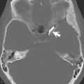
When imaging with computed tomography (CT), there is only one intrinsic contrast mechanism, and that is the electron density of the tissues being examined. Also, with CT, one does not adjust any extrinsic contrast parameters in that the kilovolts (kV) used remains high. This is not the case, however, with MRI.
There are many intrinsic contrast mechanisms that one can use in MRI. The ones discussed in this chapter include proton (spin) density (PD), T1 relaxation time, and T2 relaxation time. Images are usually acquired for which the contrast is weighted more toward one of these parameters. The key word here is weighted. Tissue contrast in the image has contributions from each of the various intrinsic contrast mechanisms, but is weighted more toward one than the others. In this context, weighting simply means the amount of contribution made to the image contrast associated with the difference between tissues on the basis of the parameter of interest (PD, T1, or T2). This weighting is accomplished by the selection of the timing parameters of the pulse sequence (set prior to scan acquisition). For spin echo sequences, these are the TR (repetition time) and the TE (echo time).
Stay updated, free articles. Join our Telegram channel

Full access? Get Clinical Tree


