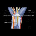
Tarsus
IMAGING ANATOMY
Overview
Arches of Foot
Bony Anatomy
 Roughly cuboidal shape
 1 ossification center: Ossifies between 9th fetal month and 6 months of age
 Articulates with calcaneus, navicular, 3rd cuneiform, and 4th and 5th metatarsals, rarely head of talus
 Sulcus at lateral margin, under which passes peroneus longus tendon
 5th metatarsal base extends beyond lateral margin
 Curved shape, concave proximally and convex distally
 1 ossification center: Ossifies in 3rd year of life
 Articulates with talus, cuboid, cuneiforms
 Large median eminence for attachment of posterior tibial tendon is located more plantar than main body of navicular
Musculature
 1st layer: Abductor hallucis, flexor digitorum brevis, abductor digiti minimi
 2nd layer: Quadratus plantae (flexor accessorius), flexor digitorum and hallucis longus tendons, lumbricals
 3rd layer: Flexor hallucis brevis, adductor hallucis, flexor digiti minimi brevis, tibialis posterior tendon
 4th layer: Plantar interossei (3), dorsal interossei (4)
 Peroneus longus tendon courses across all layers, from superficial plantar laterally to deep plantar medially


