Acquisitions Editor: Brian Brown
Product Manager: Ryan Shaw
Vendor Manager: Alicia Jackson
Senior Manufacturing Manager: Benjamin Rivera
Senior Marketing Manager: Angela Panetta
Design Coordinator: Steve Druding
Production Service: Cadmus
© 2010 by LIPPINCOTT WILLIAMS & WILKINS, a WOLTERS KLUWER business
530 Walnut Street
Philadelphia, PA 19106 USA
LWW.com
All rights reserved. This book is protected by copyright. No part of this book may be reproduced in any form by any means, including photocopying, or utilized by any information storage and retrieval system without written permission from the copyright owner, except for brief quotations embodied in critical articles and reviews. Materials appearing in this book prepared by individuals as part of their official duties as U.S. government employees are not covered by the above-mentioned copyright.
Printed in China
Library of Congress Cataloging-in-Publication Data
9780781753135
0781753135
MRI of upper extremity : shoulder, elbow, wrist, and hand / editors Lynne S. Steinbach, Christine B. Chung.
p.;cm.
Includes bibliographical references and index.
ISBN 978-0-7817-5313-5
1. Arm—Magnetic resonance imaging. I. Steinbach, Lynne S. II. Chung, Christine B.
[DNLM: 1. Upper Extremity—physiopathology. 2. Magnetic Resonance Imaging—methods. 3. Upper Extremity—anatomy & histology. WE 805 M939 2010]
RC951.M763 2010
616.7’107548—dc22
2009024490
Care has been taken to confirm the accuracy of the information presented and to describe generally accepted practices. However, the authors, editors, and publisher are not responsible for errors or omissions or for any consequences from application of the information in this book and make no warranty, expressed or implied, with respect to the currency, completeness, or accuracy of the contents of the publication. Application of the information in a particular situation remains the professional responsibility of the practitioner.
The authors, editors, and publisher have exerted every effort to ensure that drug selection and dosage set forth in this text are in accordance with current recommendations and practice at the time of publication. However, in view of ongoing research, changes in government regulations, and the constant flow of information relating to drug therapy and drug reactions, the reader is urged to check the package insert for each drug for any change in indications and dosage and for added warnings and precautions. This is particularly important when the recommended agent is a new or infrequently employed drug.
Some drugs and medical devices presented in the publication have Food and Drug Administration (FDA) clearance for limited use in restricted research settings. It is the responsibility of the health care provider to ascertain the FDA status of each drug or device planned for use in their clinical practice.
To purchase additional copies of this book, call our customer service department at (800) 638-3030 or fax orders to (301) 223-2320. International customers should call (301) 223-2300.
Visit Lippincott Williams & Wilkins on the Internet: at LWW.com. Lippincott Williams & Wilkins customer service representatives are available from 8:30 am to 6 pm, EST.
10 9 8 7 6 5 4 3 2 1
DEDICATION
To my husband, Jerry, and daughter, Sophia, for their unending moral support and unrivalled understanding. My glass overflows…
– Christine B. Chung, MD
In memory of my father, Howard L. Steinbach, M.D. who was a radiologists’ radiologist and a great role model and with special love and thanks to my husband, Eric Tepper, M.D. and son, Mark Tepper, for being devoted, accommodating, and encouraging.
– Lynne S. Steinbach, MD
CONTRIBUTORS
Rodrigo Aguiar, MD, PhD
Substitute Professor
Department of Radiology
Federal University of Paraná
Curitiba, Paraná
Radiologist
Musculoskeletal Division
DAPI – Diagnóstico Avançado Por Imagem
Curitiba, Paraná
Christina R. Allen, MD
Orthopedic Surgeon
University of California, San Francisco Medical Center
San Francisco, CA
McPherson S. Beall III, MD
Physician
Orthopedic & Fracture Clinic
Portland, OR
Luis S. Beltran, MD
Fellow
Department of Radiology, Musculoskeletal Division
New York University Langone Medical Center
New York, NY
Fellow
Department of Musculoskeletal Radiology
Hospital for Joint Diseases
New York, NY
John P. Belzer
Assistant Clinical Professor
Department of Orthopaedics
University of California, San Francisco
San Francisco, CA
Vice Chairman
Department of Orthopedics
California Pacific Medical Center
San Francisco, CA
Frederic W. Bost
Assistant Clinical Professor
Department of Orthopaedics
University of California, San Francisco
San Francisco, CA
Attending Staff
Department of Orthopedic Surgery
California Pacific Medical Center
San Francisco, CA
Linda Chen, MD
Assistant Professor
Department of Radiology
UCSD Medical Center and VASDHS
San Diego, CA
Christine B. Chung, MD
Associate Professor
Department of Radiology
University of California, San Diego
San Diego, CA
Edward Diao, MD
Professor Emeritus
Department of Orthopaedic Surgery
Founder and Former Chief
Division of Hand, Upper Extremity and Microvascular Surgery
Department of Orthopaedic Surgery and Neurosurgery
University of California, San Francisco
San Francisco, CA
Chief of Hand Surgery
Department of Orthopaedic Surgery
California Pacific Medical Center
San Francisco, CA
Tudor Hesketh Hughes, MD, FRCR
Professor of Radiology
Department of Radiology
University of California, San Diego Medical Center
San Diego, CA
David Malfair, MD
Clinical Instructor
Department of Radiology
University of British Columbia
Vancouver, BC
Department of Radiology
Vancouver General Hospital
Vancouver, BC
Aurea V.R. Mohana-Borges, MD
Assistant Professor of Radiology
Carlos Chagas Postgraduate Medical Institute (IPMCC)
Staff Radiology
Hospital de Clinicas de Niteroi (HCN)
Hospital São Lucas (HSL) and ProEcho Niteroi
São Lucas, Brazil
William B. Morrison, MD
Professor
Department of Radiology
Thomas Jefferson University
Philadelphia, PA
Director, Division of Musculoskeletal and General Diagnostic Radiology
Department of Radiology
Thomas Jefferson University Hospital
Philadelphia, PA
Geoffrey Riley, MD
Assistant Clinical Professor
Department of Radiology
University of California, San Francisco
San Francisco, CA
Marc R. Safran, MD
Professor
Department of Orthopaedic Surgery
Associate Director
Department of Sports Medicine
Stanford University
Redwood City, CA
Lynne S. Steinbach, MD
Professor of Radiology and Orthopaedic Surgery
University of California, San Francisco
San Francisco, CA
Hiroshi Yoshioka, MD, PhD
Associate Professor
Department of Radiology
Harvard Medical School
Cambridge, MA
Director, Musculoskeletal CT and MRI
Department of Radiology
Brigham and Women’s Hospital
Boston, MA
FOREWORD
I find myself both honored and privileged to have the opportunity to write the Foreword for this book. Beyond that, I am proud to have played some role in the academic development of these two authors who are now internationally recognized authorities in the field of musculoskeletal imaging. With the publication of this text, they have accomplished far more than the collection and presentation of state of the art information dealing with MR imaging of the upper extremity. Rather, Christine Chung and Lynne Steinbach provide us with a blockbuster of a publication. A blockbuster refers to something that is forcefully or overwhelmingly impressive, effective or influential, and I cannot think of a more appropriate choice for a single word that describes what is presented here. That the book is impressive and effective and will become influential should surprise no one, certainly not me, if one is familiar with what these two musculoskeletal radiologists have achieved through the years leading up to this publication. They both have a proven track record of thoughtful and original scientific investigation, but many other authors have this attribute. They both are well respected clinical radiologists, possessing extraordinary diagnostic skills, but then again there are other authors who have such skills. And both Christine and Lynne possess impressive knowledge not just in the realm of diagnostic imaging but also with regard to orthopaedics, sports medicine, and rheumatology … but there are a handful of other authors who have similar credentials.
No, what distinguishes these two authors from most others relates to their dedication to teaching and their effectiveness as educators. To write a blockbuster requires that the available information be critically assessed, teased and, in some instances, discarded such that the essentials remain. Next, to be effective, the authors must present this remaining material in a highly organized and disciplined fashion. Finally, they must supplement this presentation through the judicious use of images, drawings, and tables. To do all this requires the attention, dedication and energetic input of accomplished teachers—and I can think of no better persons to do this than Christine and Lynne. Any one who has had the opportunity to listen to these two educators at the podium with a large audience in attendance or around the computer console with only a few nearby is well aware of their unique abilities as educators. They present material with an order and cadence of words that bespeak years of experience and this cadence echoes in the pages of this book.
Despite the complexity of the material presented in this text and the complete range of clinical problems that are addressed (and they are all here), this might be classified as an “easy read”. And there-in rests the true beauty of this book. For at the end, the reader who has taken the time to digest this material comes away from the experience with the satisfaction that he or she has learned a great deal and will be a better physician for doing so. That person will have learned the detailed imaging findings that accompany rotator cuff disorders and instability of the shoulder, ligamentous and tendinous alterations about the elbow, abnormalities of the triangular fibrocartilage and carpal tunnel of the wrist, finger malalignments and deviations, and many more subjects too numerous to list here. Furthermore, the reader will have learned all this without becoming fatigued and will bring new energy to his or her clinical practice similar in intensity to the energy and enthusiasm that can be found in each of the pages of this text.
Make no mistake about this. This is a blockbuster of a publication. It belongs on the shelf and in the hands of all radiologists who are involved in the interpretation of musculoskeletal imaging studies. Orthopaedic surgeons, rheumatologists and sports medicine physicians whose practice includes the care of patients with clinical problems of the upper extremity also would be well advised to purchase this book. And for the residents and fellows who have an interest in or who are required to know the principles of such musculoskeletal imaging, I would suggest that this would be a wise investment.
As I indicated at the outset, I am deeply honored, privileged and proud to have been asked to write this Foreword.
Donald Resnick, MD
PREFACE
At its initial inception this project was intended to expand upon the work of Steinbach, et al, in Shoulder Magnetic Resonance Imaging, broadening the focus to include an articular-based approach to MR imaging in the upper extremity. To a large degree, the decision to include the elbow, wrist and fingers within the scope of this work was anchored in the realization that the historical underrepresentation of these exams within our clinical practices has changed remarkably in the recent past. The demand for technically sound MR imaging studies, accurate diagnoses and detailed characterization of pathology in these regions has increased and obligates the radiologist to commensurately establish and or expand their familiarity with these joints.
This text begins with an atlas that correlates MR imaging, and in some cases MR arthrography, with sectional anatomic specimen photographs and emphasizes a theme that has been well-established by Dr. Donald Resnick, the guru of musculoskeletal radiology and an invaluable mentor to both authors. This theme, of course, is that one must have a detailed understanding of normal anatomy before pathology can be detected and characterized. “Yet, the radiograph is but a mirror and its image a reflection of the underlying anatomy and pathology” according to Donald Resnick in his preface to Diagnosis of Bone and Joint Disorders, First Edition. In the setting of MR imaging, perhaps the mirror has become a window through which we look into joints and even tissues to characterize anatomy and subsequently pathology. The production of this atlas was a labor of the love of anatomy for the authors and illustrator of this text, and a labor that reaped the benefit of the ownership of this knowledge. We consider this an empowering benefit, and one that we hope you will share with us.
While this is an MR imaging text that highlights the basics as well as the most up-to-date concepts regarding the upper extremity joints, the reader will frequently encounter radiographic and computed tomographic correlation, as well as dialogue with respect to the role of MR imaging versus other imaging methods in the evaluation of pathologic entities. Both authors recognize the importance of MR imaging in our musculoskeletal practices, but in no way want to lessen the impact and importance of radiographic evaluation. While the interpretation of radiographs is not considered as glamorous as MR imaging evaluation, the authors maintain that the former requires more expertise when done well, and offers invaluable additional information when interpreting MR imaging studies. Indeed, we often advise our residents that MR imaging can be an important tool to enhance plain film interpretation!
While the majority of this text has been written by the editors, the contributing authors and the illustrator of the text have provided the highest level of expertise and quality of information. The process of writing this book and editing the contributions of our colleagues was an educational experience that broadly expanded our knowledge base. We hope that this text will become not only an important reference, but provide a valuable foundation from which the reader can base their clinical practice and further intellectual discovery.
ACKNOWLEDGEMENTS
The process of writing this textbook has provided me with the reaffirmation that intellectual stimulation and learning is a driving force in my life, one that provides me with immense personal and professional satisfaction. To this end, I am indebted to both my co-editor and the contributors of this text, who have provided educational material of an unparalleled quality. Further I must recognize the residents, fellows and research fellows that have inspired me, through intuitive and often perplexing questions, to strive to find better and various ways to educate and to promote individual discovery—for this is the very essence of the academic radiologist. I must also recognize the supportive environment of my early mentor, Dr. Joseph Yu, my subsequent academic partners, particularly Drs. Donald Resnick and Mini Pathria, and the co-editor of this textbook, Dr. Lynne Steinbach, whose sheer and unadulterated love of musculoskeletal radiology was, and still is, a motivating factor in my career.
While the contributions of several people are acknowledged in the figures, those of two people stand out, Drs. Rama Sharma and Eric Smith. Indeed, their contributions were so numerous that they have been deemed ‘major contributors’ to this text, and their names have not been noted in each and every figure they provided. To them, I would like to extend my sincere thanks. A special thank you must also be extended to the illustrator of this textbook and co-author of the atlas, Dr. Aurea Mohana-Borges. As you will see, the detail and quality of the illustrations she has provided richly enhance the textbook in an invaluable way. Similarly, Debra Trudell deserves acknowledgement for her help in preparation of the MR imaging atlas.
Ryan Shaw and Ruth Einstein proved to be the coordinating forces that moved this textbook to its final stages of completion—a task that deserves special thanks as this is often the point where the process begins to lose momentum. My research laboratory manager, Sheronda Statum deserves a hearty thank you for her support in all things pertaining to the production of this book, though well outside the auspices of her official responsibilities. Finally, I must acknowledge the support of Dr. Jerry Dwek. It is the minority of authors that can thank his or her spouse not only for their patience, understanding, and moral support—but additionally for their intellectual contributions relating to the subject matter. This is the added advantage of having a pediatric and musculoskeletal trained radiologist combined with wonderful husband.
– Christine B. Chung, MD
I am indebted to the inspiration of Dr. Christine Chung in co-authoring the subjects in this book. Her strength, intelligence, and enthusiasm are unmatched. Additionally, her persistence ensured the successful completion of this project. I would also like to thank Dr. Aurea Mohana-Borges, a musculoskeletal radiologist and great artist with a wonderful talent for crystallizing concepts through diagrams that are simple and understandable. Gratitude goes to several authors of various chapters, including Drs. Geoff Riley, Dave Malfair, Hiroshi Yoshioka, Lina Chen, and William Morrison and their co-authors. Kudos to several of my orthopedic colleagues, including Drs. Christina Allen, Edward Diao, and John Belzer and their co-authors who present excellent clinical correlations. The current editor, Ryan Shaw, made completion of the text efficient and fulfilling. Drs. Suzanne Anderson and Vincent McCormick provided moral support during this period, while contributing several images. Special thanks go to Dr. Donald Resnick for keeping me on my toes when it comes to the presentation of new concepts in musculoskeletal imaging and for his teaching, mentoring, and supportive encouragement throughout my career.
– Lynne S. Steinbach, MD
CONTENTS
1 Anatomy of Upper Extremity Joints with Cadaveric Correlation
2 Technical Considerations for MRI of Upper Extremity Joints
3 Clinical Evaluation of the Painful Shoulder
5 MRI of Glenohumeral Instability
6 Long Bicipital Tendon Including Superior Labral Anterior–Posterior Lesions
7 Miscellaneous Conditions of the Shoulder
Luis S. Beltran, William B. Morrison and Aurea V. R. Mohana-Borges
9 Clinical Evaluation of the Painful Elbow
10 Elbow Ligaments and Instability
11 Elbow Tendons and Epicondylitis
13 Miscellaneous Disorders of the Elbow
14 Clinical Evaluation of the Painful Wrist and Hand
15 Wrist Ligaments and the Triangular Fibrocartilage Complex
16 The Carpal Tunnel and Guyon’s Canal
17 Miscellaneous Conditions of the Wrist
20 Miscellaneous Disorders of the Finger
Shoulder
Axial
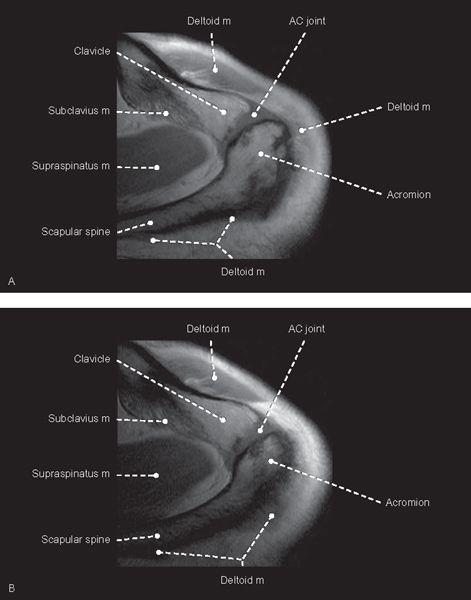
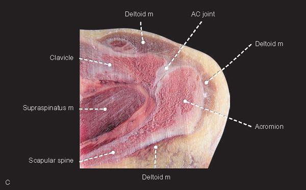
FIGURE 1.1
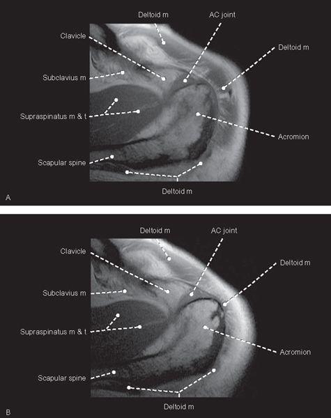
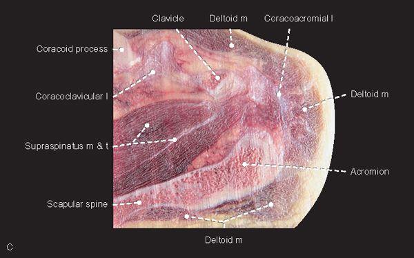
FIGURE 1.2
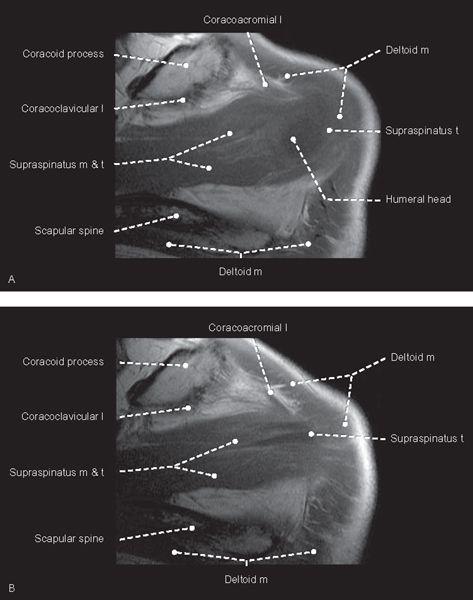
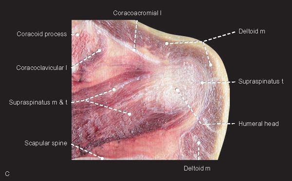
FIGURE 1.3
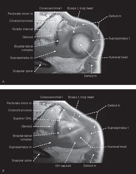
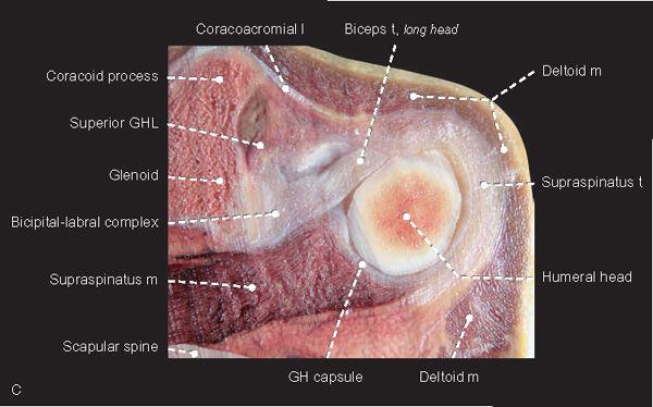
FIGURE 1.4
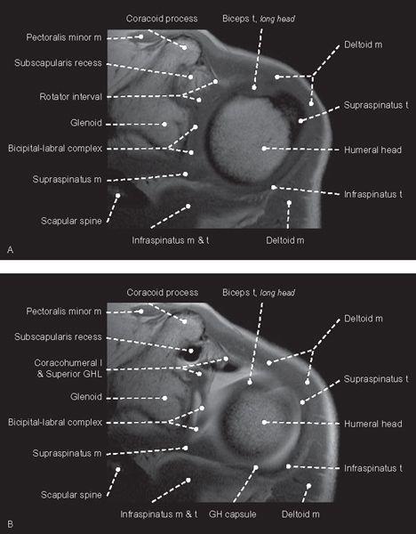
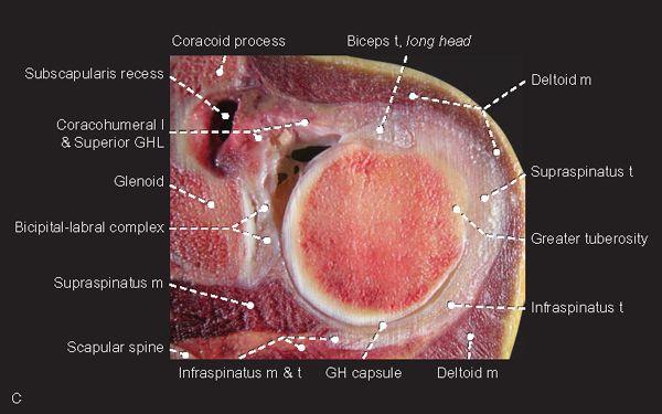
FIGURE 1.5
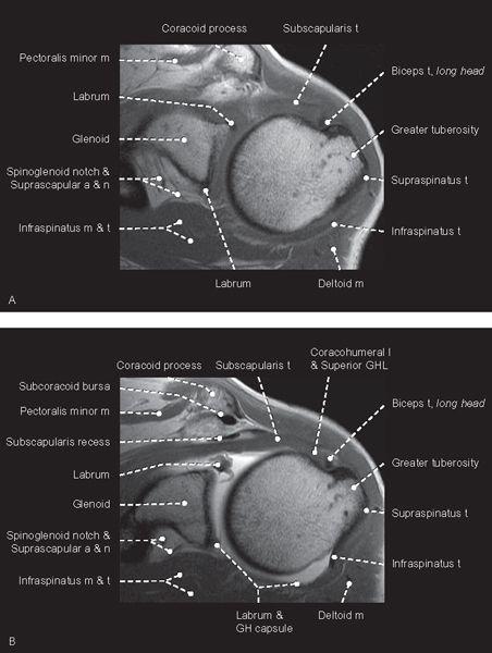
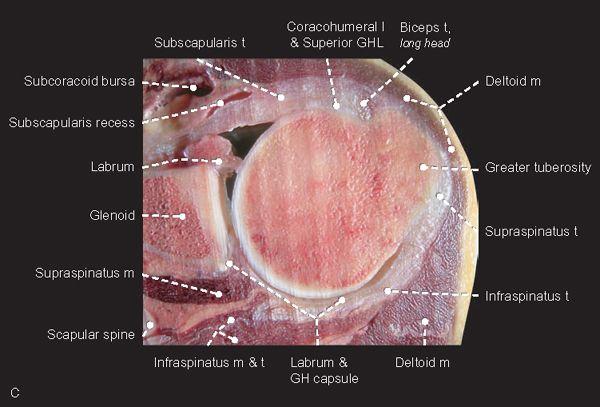
FIGURE 1.6
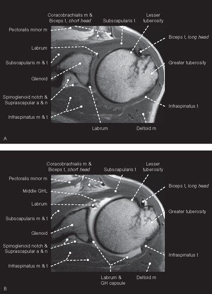
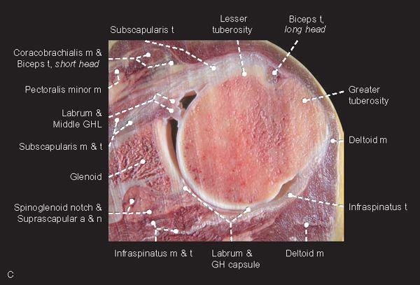
FIGURE 1.7
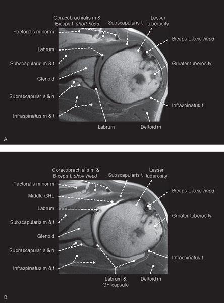
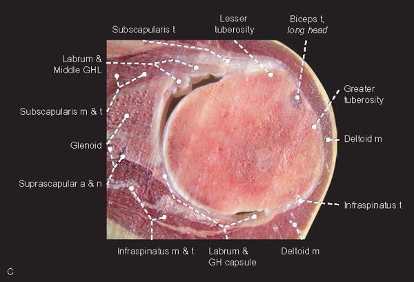
FIGURE 1.8
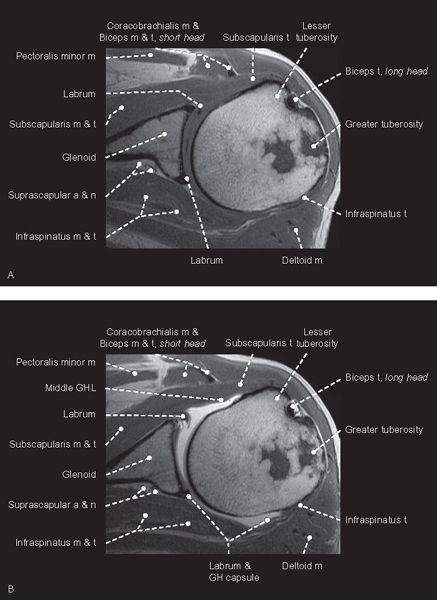

FIGURE 1.9
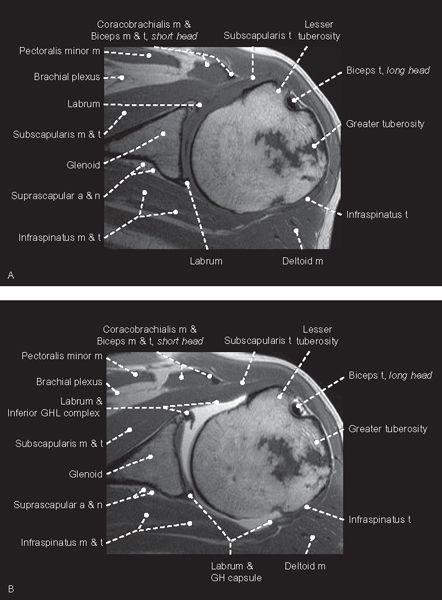
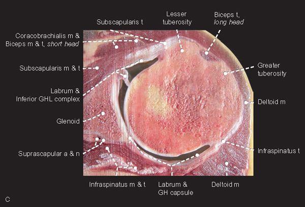
FIGURE 1.10
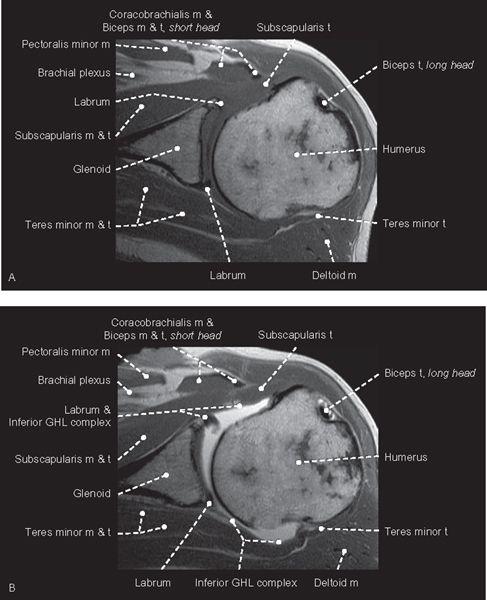
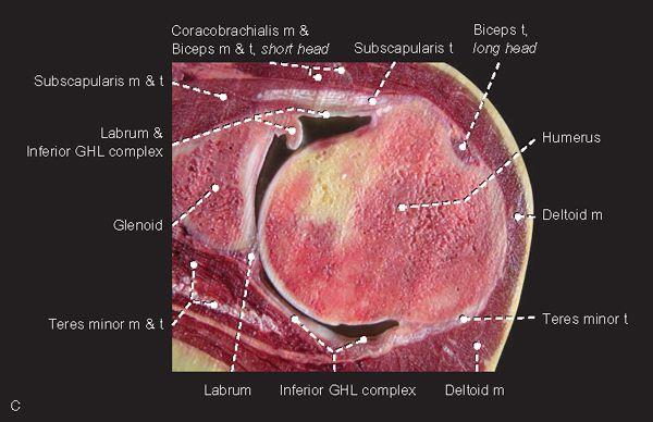
FIGURE 1.11
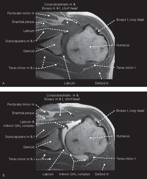
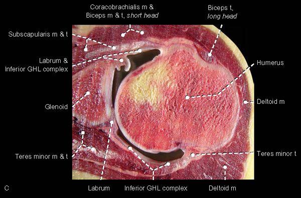
FIGURE 1.12
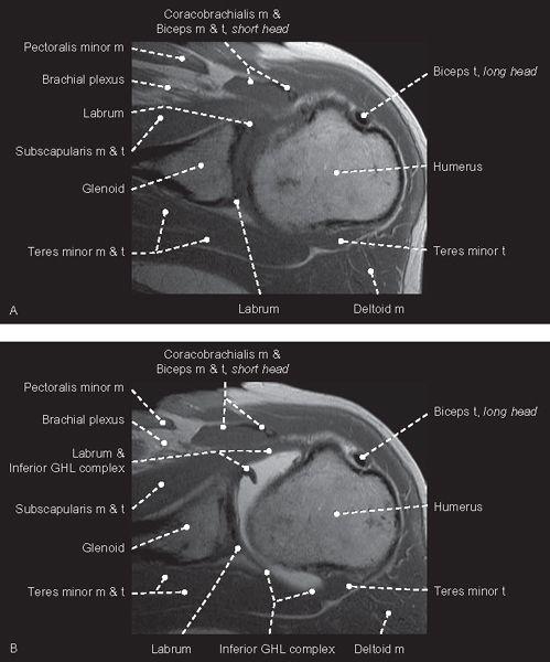
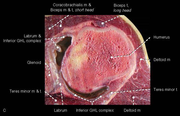
FIGURE 1.13
Coronal
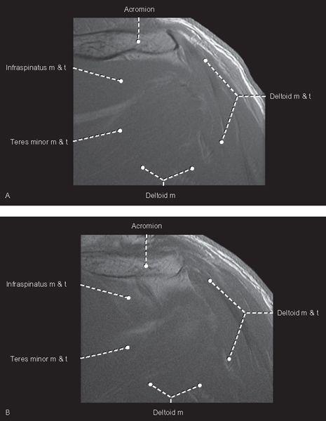
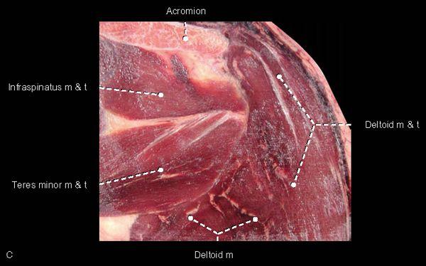
FIGURE 1.14
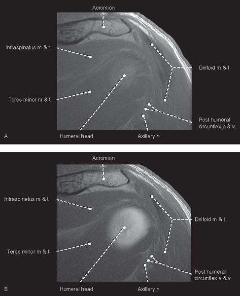
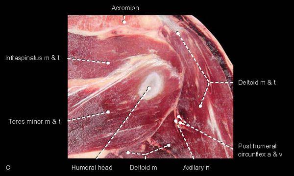
FIGURE 1.15
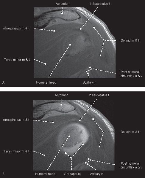
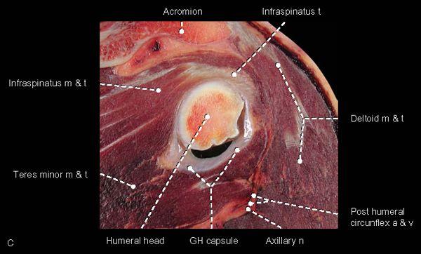
FIGURE 1.16
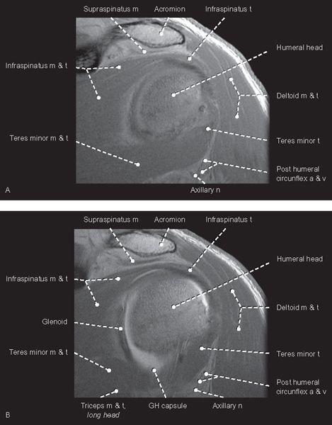
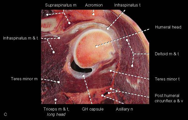
FIGURE 1.17
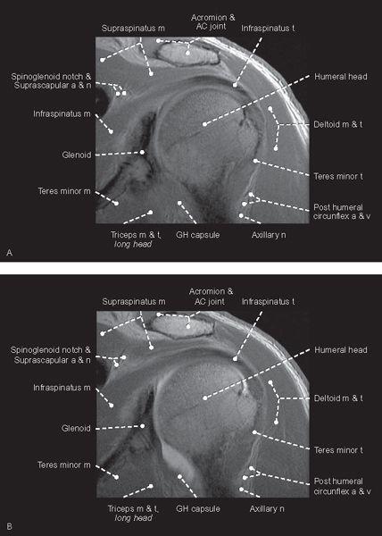
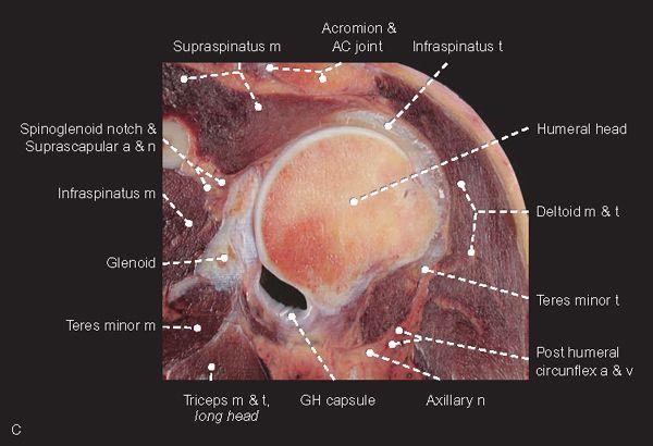
FIGURE 1.18
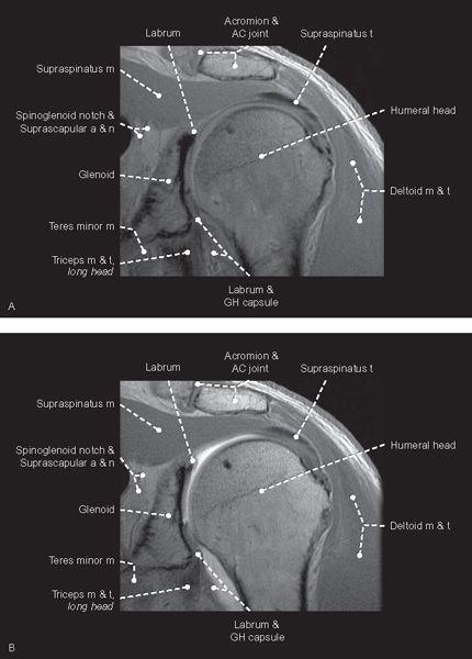
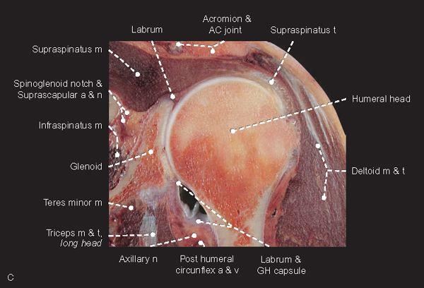
FIGURE 1.19
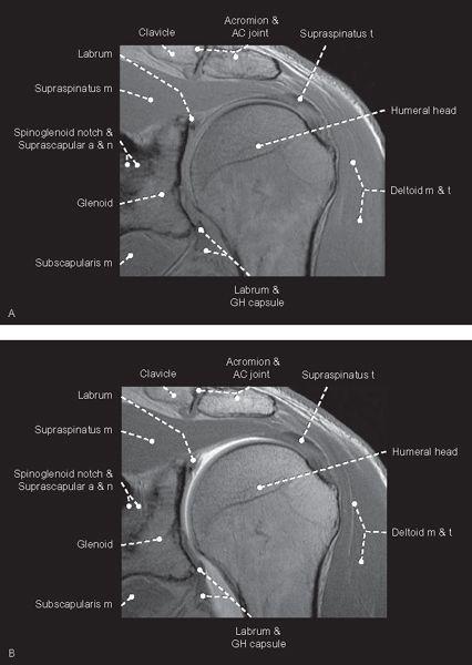
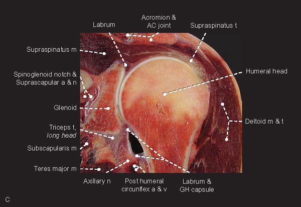
FIGURE 1.20
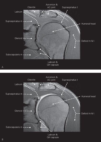
FIGURE 1.21
FIGURE 1.22
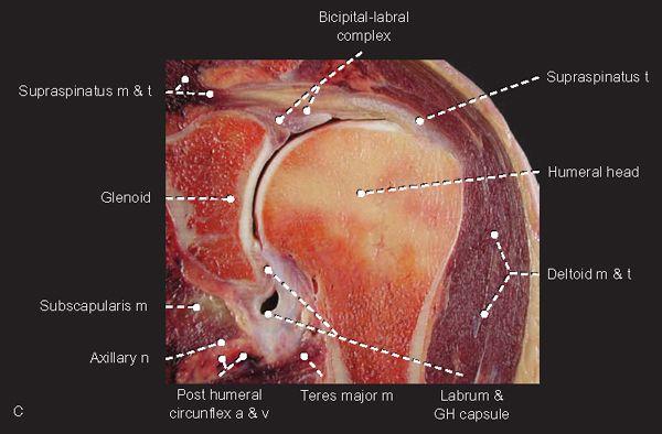
FIGURE 1.23
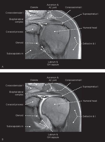
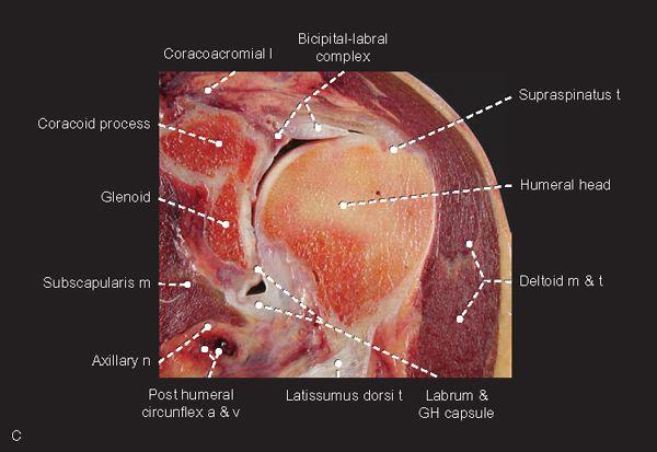
FIGURE 1.24
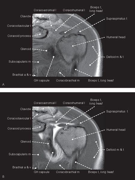
FIGURE 1.25
FIGURE 1.26
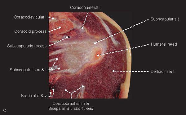
FIGURE 1.27
Sagittal
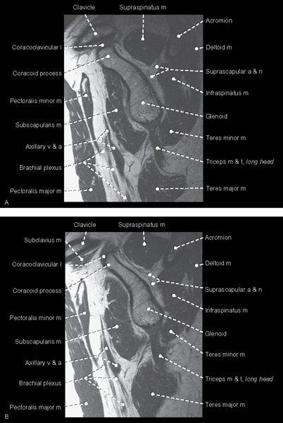
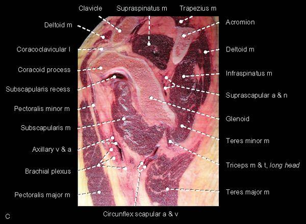
FIGURE 1.28
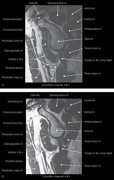
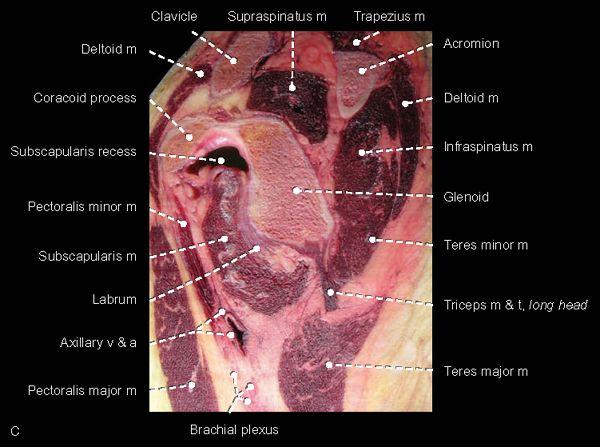
FIGURE 1.29
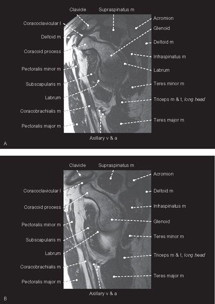
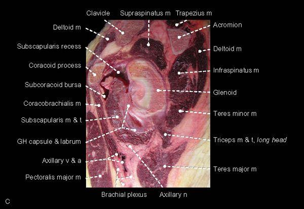
FIGURE 1.30
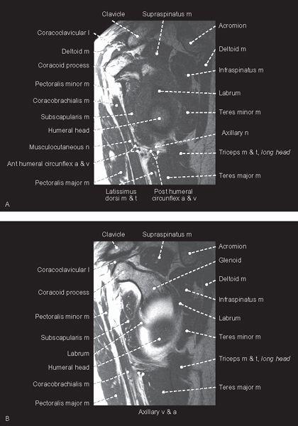
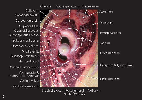
FIGURE 1.31
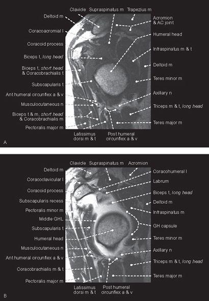
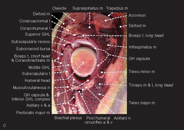
FIGURE 1.32
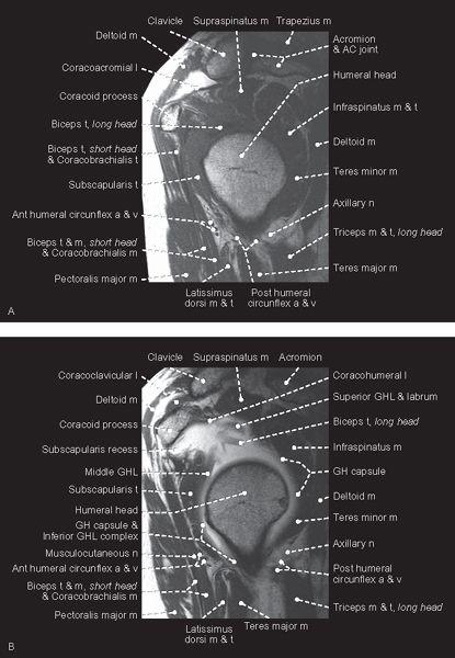
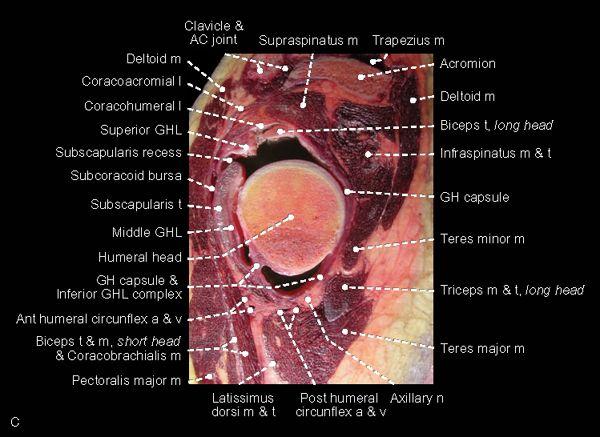
FIGURE 1.33
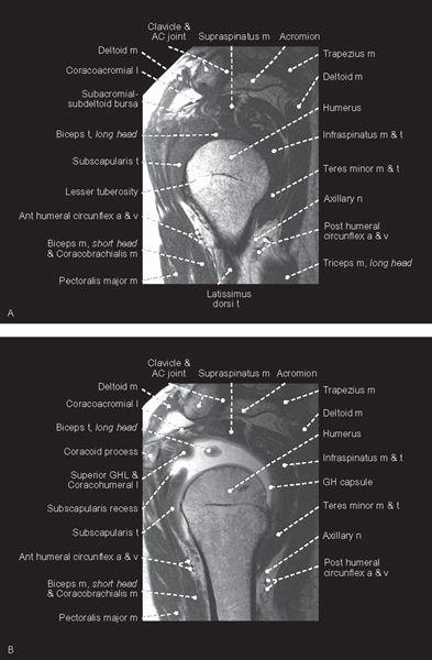
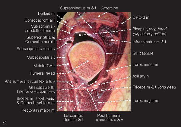
FIGURE 1.34
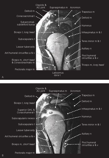
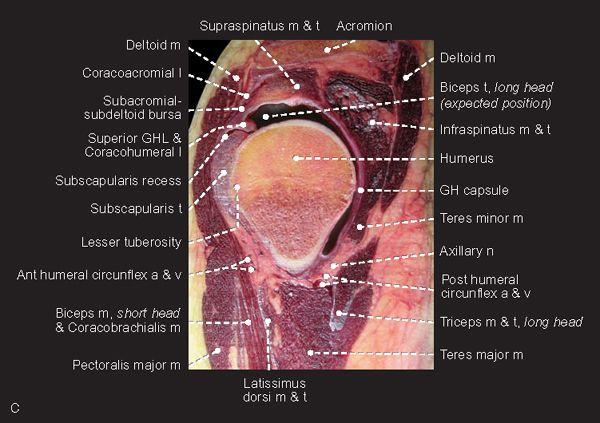
FIGURE 1.35
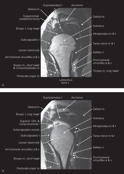

FIGURE 1.36
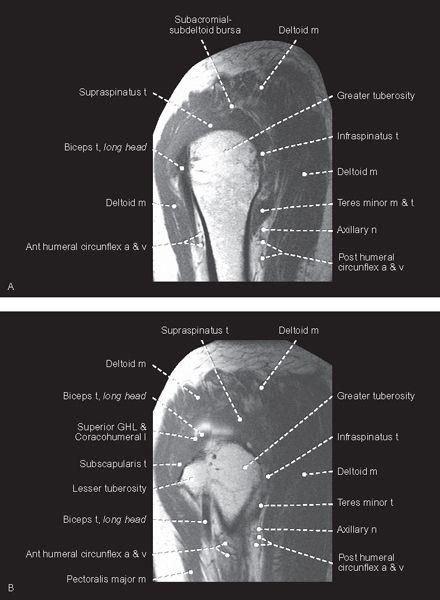
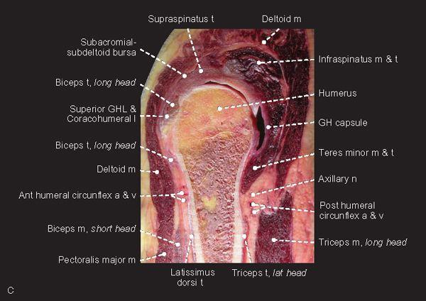
FIGURE 1.37
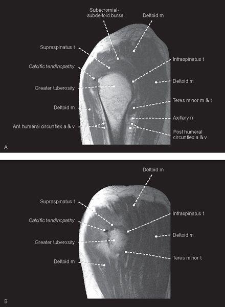
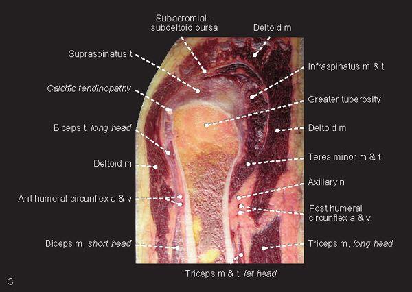
FIGURE 1.38
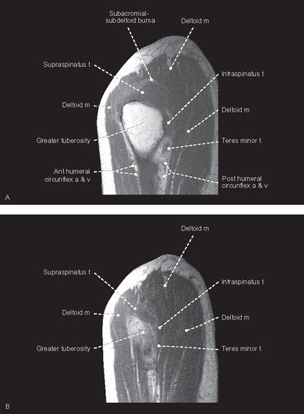
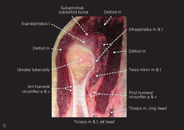
FIGURE 1.39
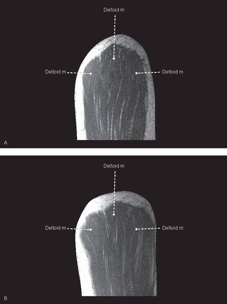
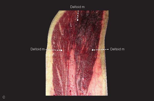
FIGURE 1.40
Elbow
Axial
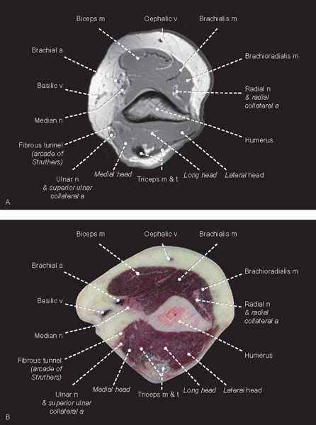
FIGURE 1.41

FIGURE 1.42
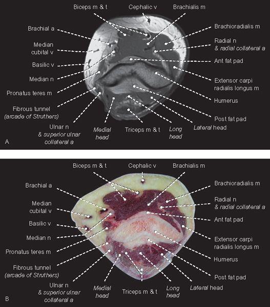
FIGURE 1.43
FIGURE 1.44
FIGURE 1.45
FIGURE 1.46
FIGURE 1.47
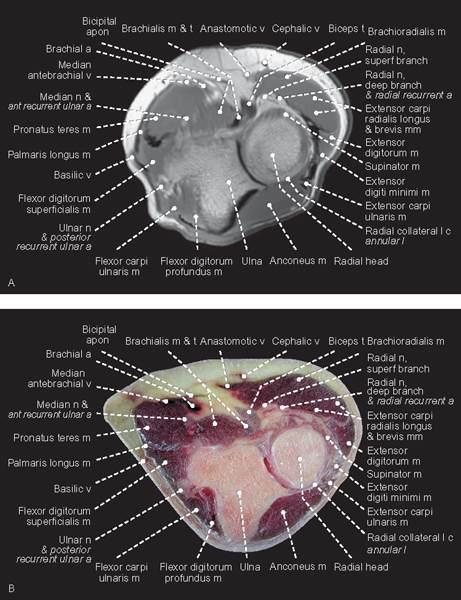
FIGURE 1.48
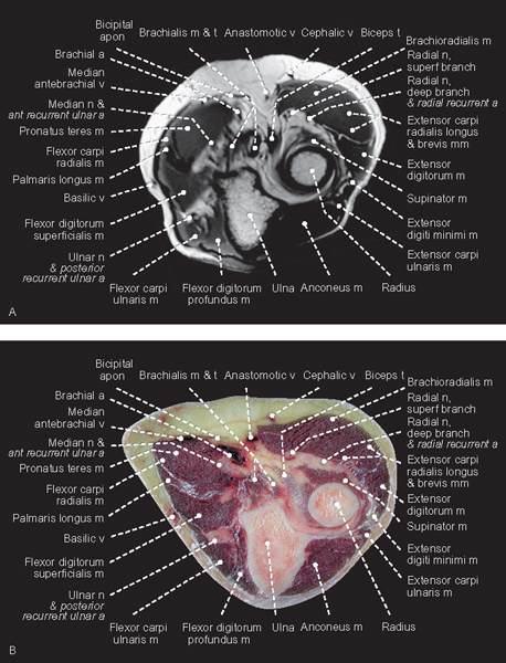
FIGURE 1.49
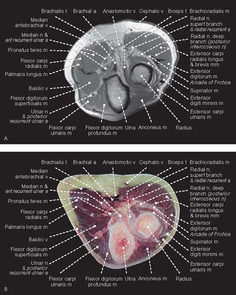
FIGURE 1.50
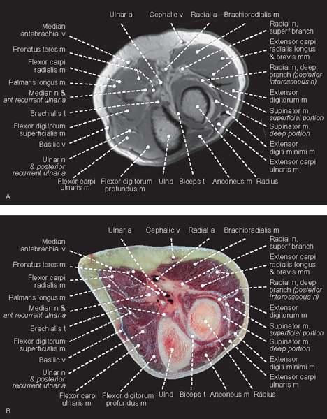
FIGURE 1.51
Coronal
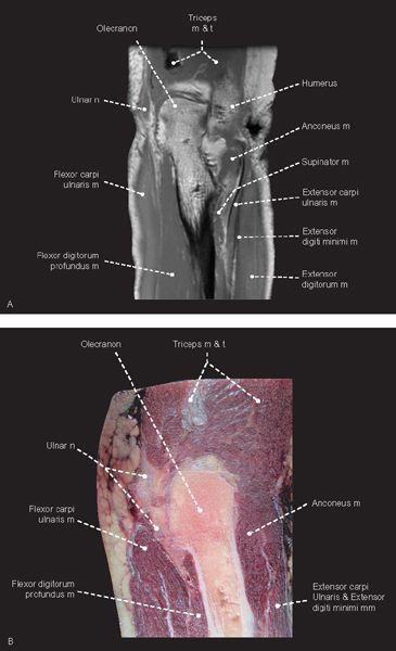
FIGURE 1.52
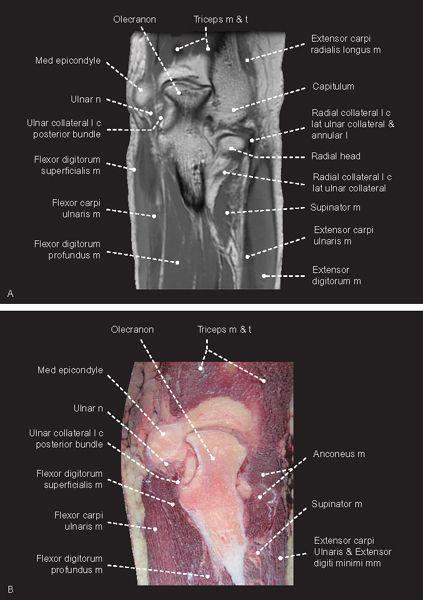
FIGURE 1.53
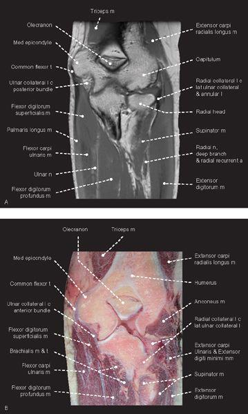
FIGURE 1.54
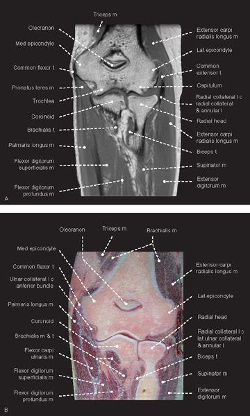
FIGURE 1.55
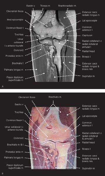
FIGURE 1.56
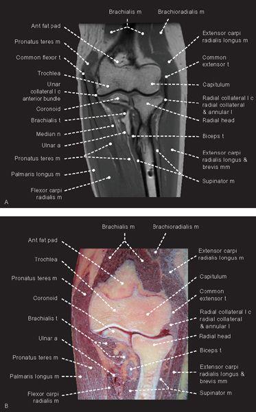
FIGURE 1.57
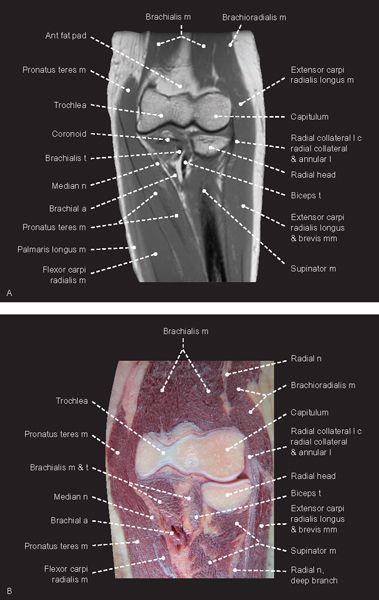
FIGURE 1.58
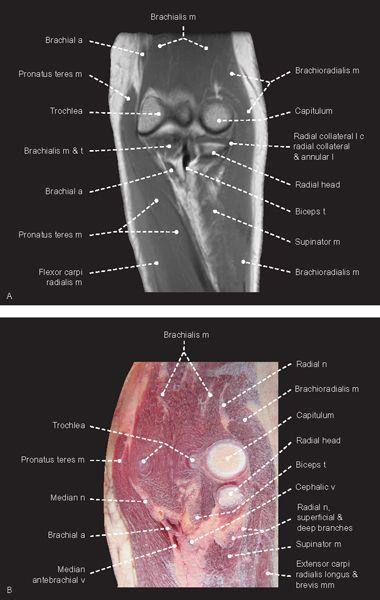
FIGURE 1.59
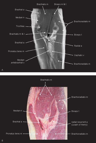
FIGURE 1.60
Sagittal
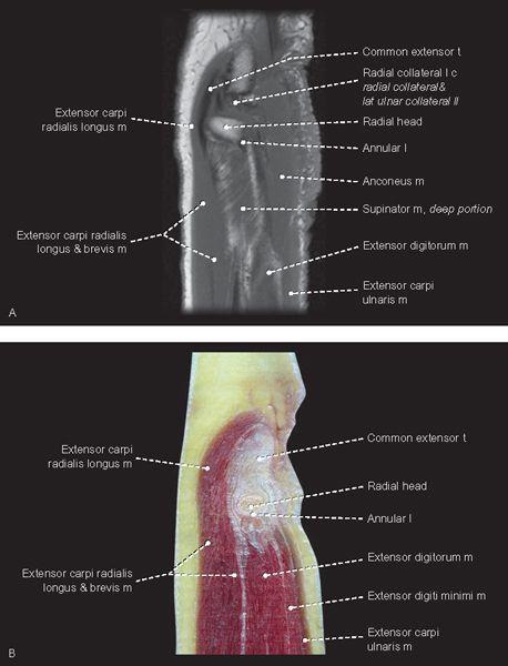
FIGURE 1.61
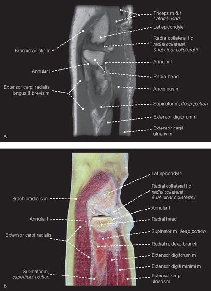
FIGURE 1.62
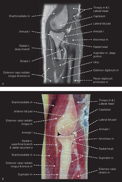
FIGURE 1.63
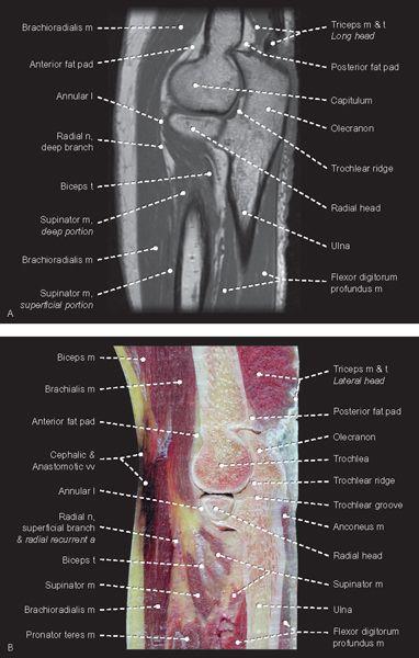
FIGURE 1.64
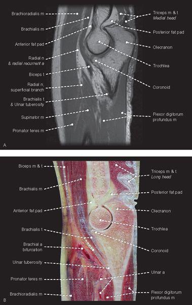
FIGURE 1.65
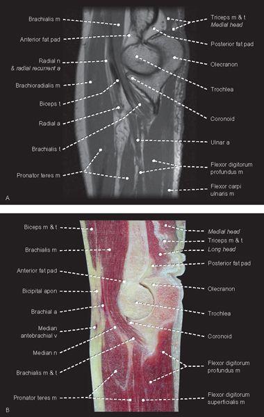
FIGURE 1.66
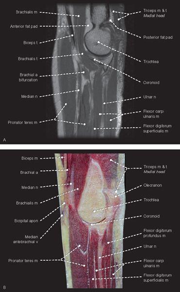
FIGURE 1.67
FIGURE 1.68
Wrist
Axial
FIGURE 1.69
FIGURE 1.70
FIGURE 1.71
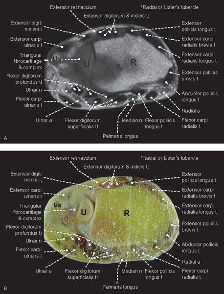
FIGURE 1.72
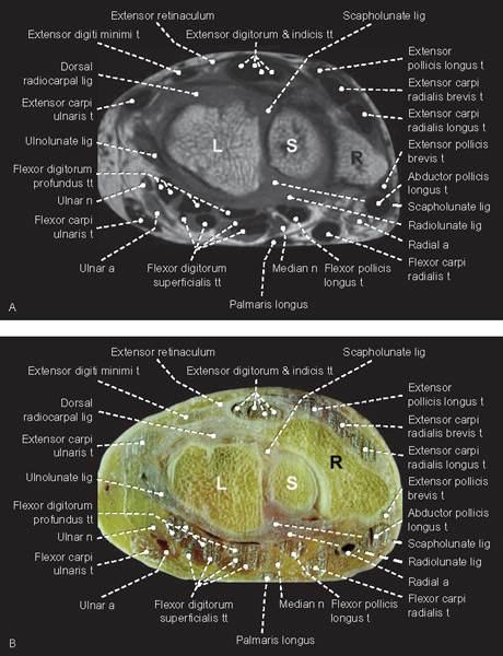
FIGURE 1.73
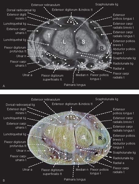
FIGURE 1.74
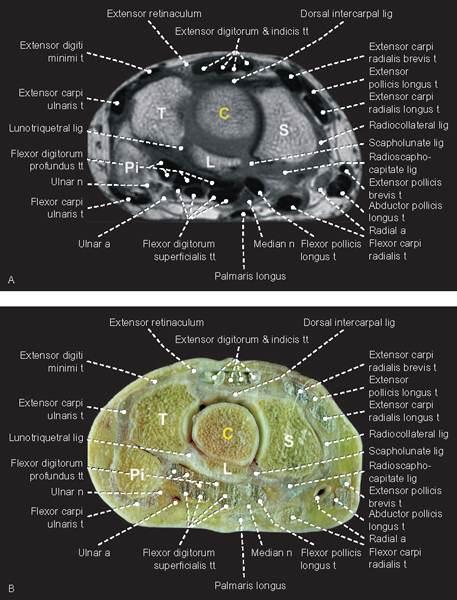
FIGURE 1.75
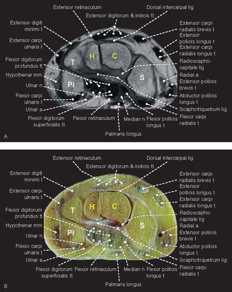
FIGURE 1.76
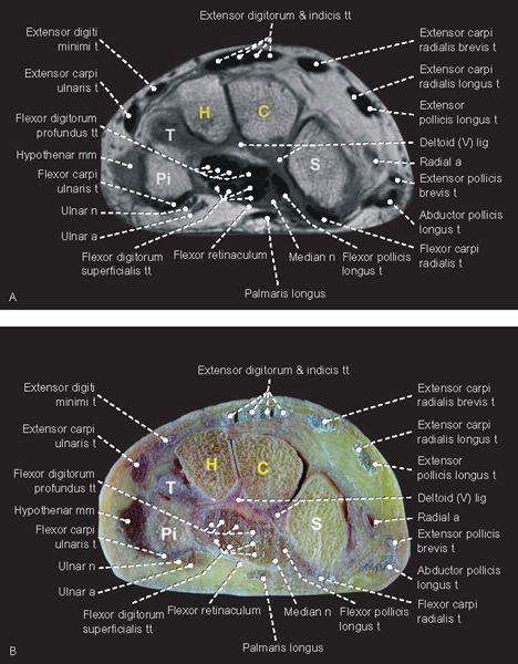
FIGURE 1.77
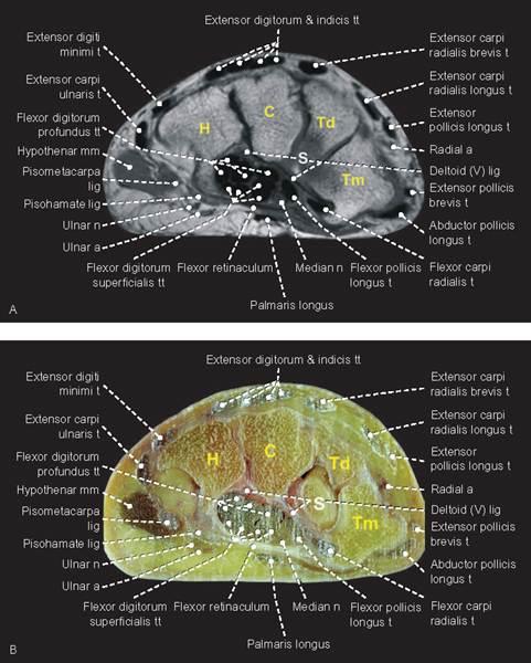
FIGURE 1.78
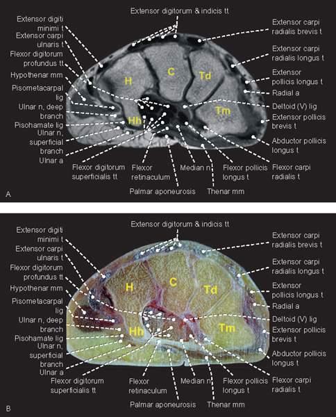
FIGURE 1.79
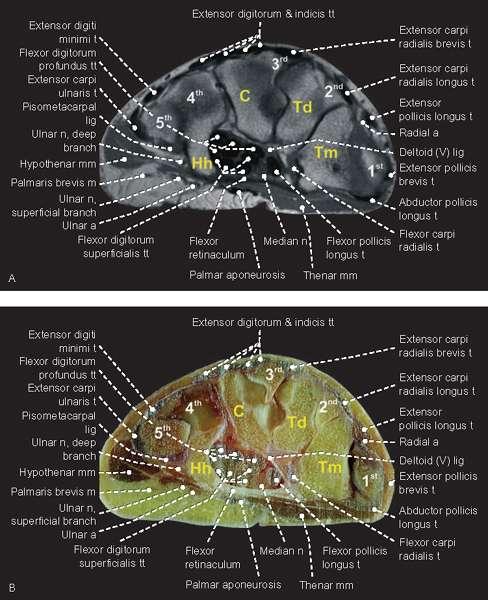
FIGURE 1.80
Coronal
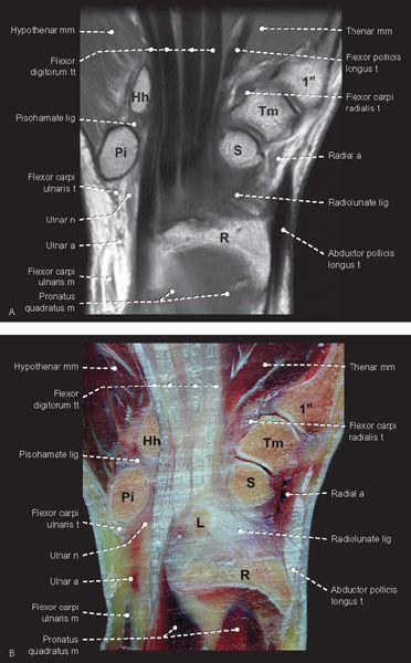
FIGURE 1.81
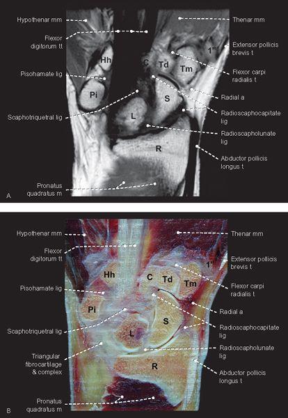
FIGURE 1.82
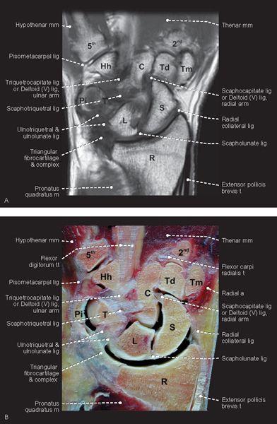
FIGURE 1.83
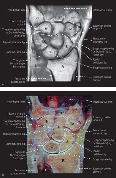
FIGURE 1.84
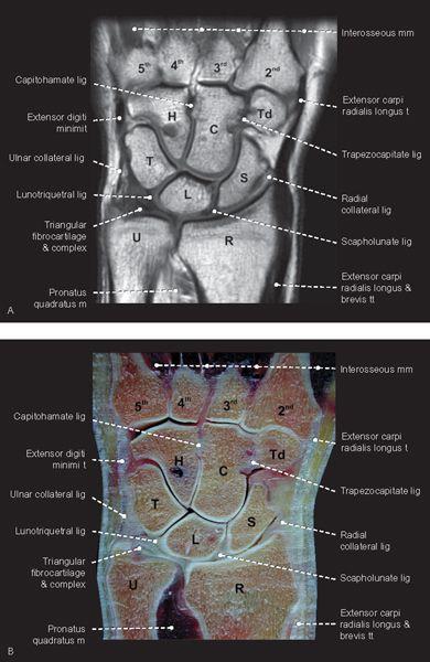
FIGURE 1.85
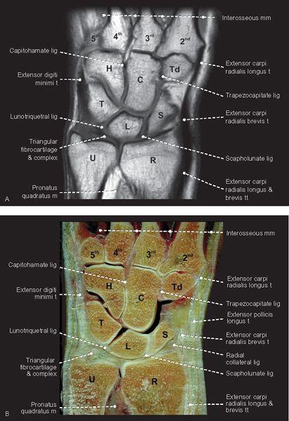
FIGURE 1.86
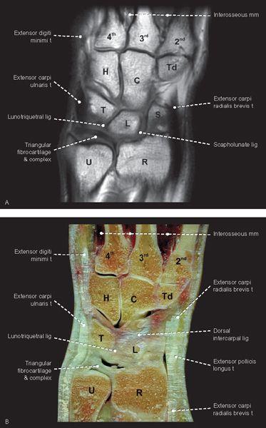
FIGURE 1.87
FIGURE 1.88
Sagittal
FIGURE 1.89
FIGURE 1.90
FIGURE 1.91
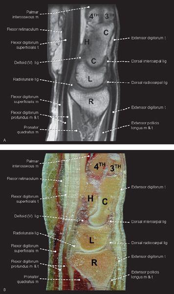
FIGURE 1.92
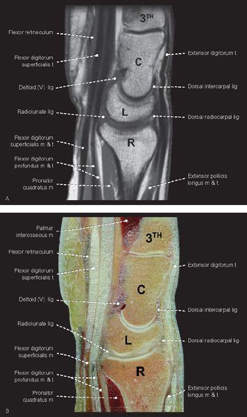
FIGURE 1.93
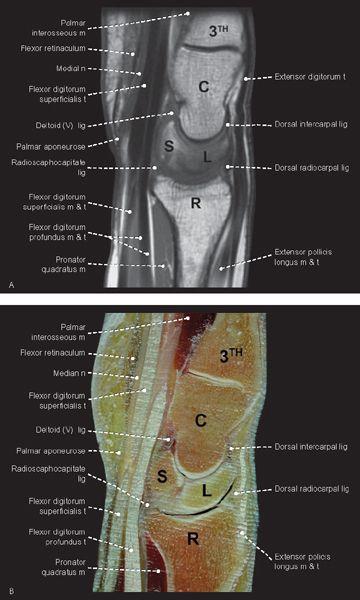
FIGURE 1.94
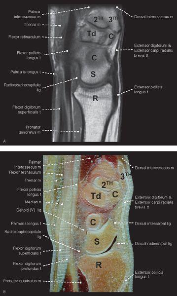
FIGURE 1.95
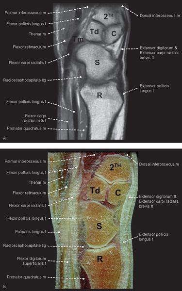
FIGURE 1.96
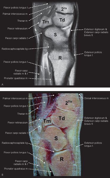
FIGURE 1.97
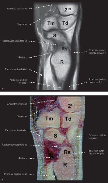
FIGURE 1.98
Finger
Axial
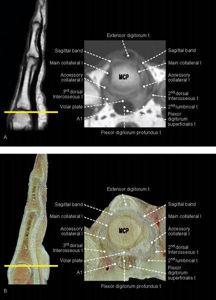
FIGURE 1.99
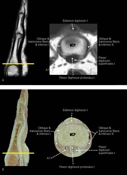
FIGURE 1.100
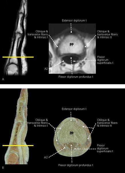
FIGURE 1.101

FIGURE 1.102
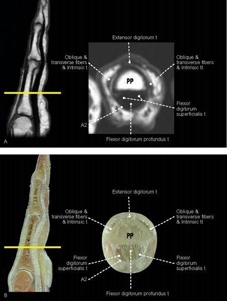
FIGURE 1.103
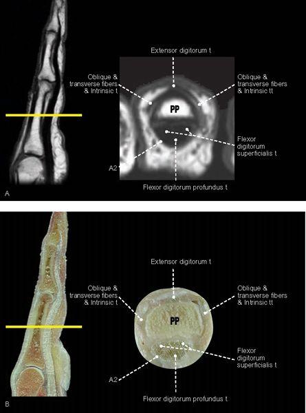
FIGURE 1.104
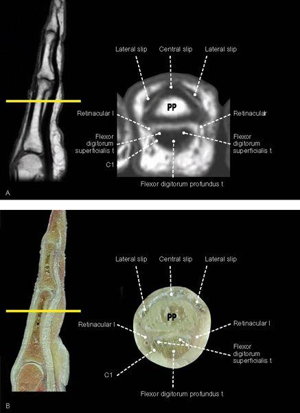
FIGURE 1.105
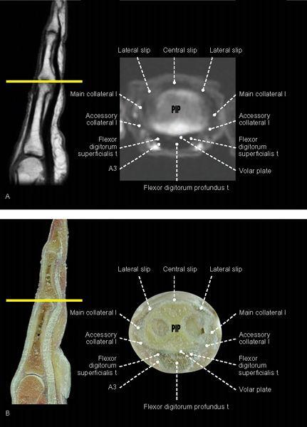
FIGURE 1.106
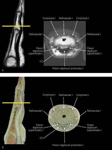
FIGURE 1.107
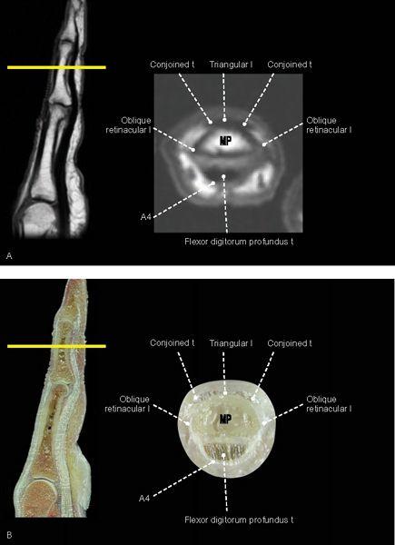
FIGURE 1.108
FIGURE 1.109
FIGURE 1.110
Coronal
FIGURE 1.111
FIGURE 1.112
FIGURE 1.113
FIGURE 1.114
FIGURE 1.115
Sagittal
FIGURE 1.116
FIGURE 1.117
FIGURE 1.118

FIGURE 1.119
FIGURE 1.120
Thumb
Axial
FIGURE 1.121
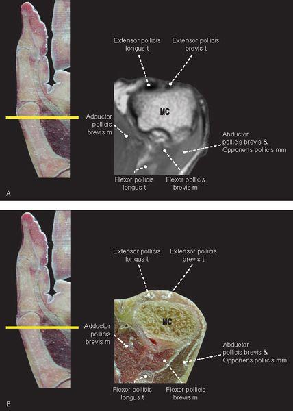
FIGURE 1.122
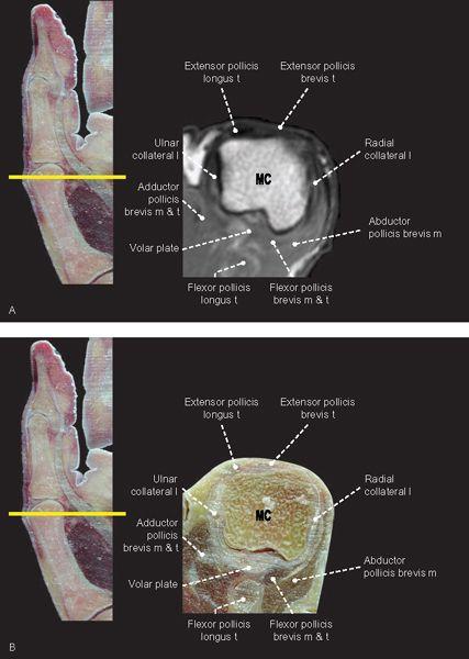
FIGURE 1.123
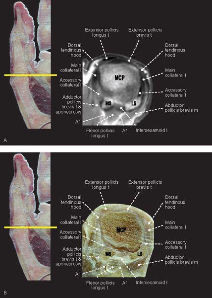
FIGURE 1.124
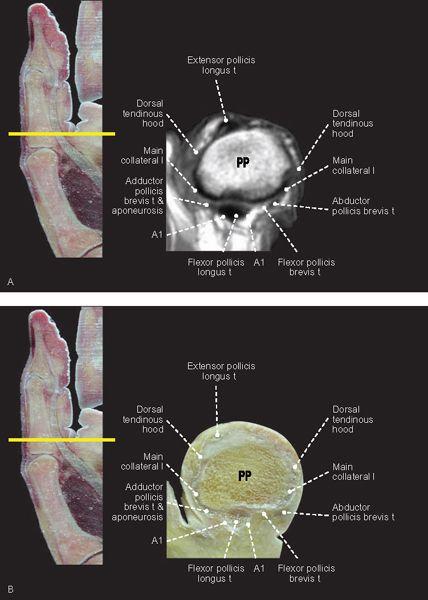
FIGURE 1.125
FIGURE 1.126
FIGURE 1.127
FIGURE 1.128
FIGURE 1.129
Related posts:

Stay updated, free articles. Join our Telegram channel

Full access? Get Clinical Tree


