Fig. 3.1
The progression in stereotactic localization from 1906 to the modern time. (a) A replicate of the horsely and clarke frame (open source from Google images). (b) A modern leskell frame (image used with permission from Elekta). (c, d) A frameless system used in the operating room where the reference fiducial system is attached to the head-rest and surface anatomical landmarks are co-registered with the preoperative CT and MRI fused images, thus allowing real-time navigation and target localization
History of Stereotaxis
The development of sterotactic localization parallels those of brain imaging, but the earliest applications of stereotaxis predate the advent of imaging modalities. Dittmar in 1873 describes a study of the medulla oblongata of cat using a device to read off the distance from fixed dependable surface anatomical landmark. Zernov in Russia used a similar approach in 1890 to locate deep structures in humans [2]. The L’encephalometer was a relatively simple device anchored onto the meridian of the human skull; it used the polar coordinate and arc-based angulation system to mark the trajectory to the designated structures of the brain. In 1889, it was used to locate the central sulcus of the human brain for the aspiration of an abscess [3]. Ultimately, its contribution to neurosurgery was overshadowed by the work of Horsely and Clarke.
Not unlike Zenov’s device, Horsely’s device was also anchored onto the skull by using a pair of ear bars. The innovation of their device was having a separate base frame and a headpiece. The base frame fixed onto the skull rigidly and established the horizontal planes along Reid’s line, thus allowing the coordinates to be read along the infraorbital border to the external auditory meatus. The headpiece was then incorporated perpendicular onto the basal frame to adjust the vertical plane. The headpiece carried a micro-manipulator that was perpendicular to the frame. It was through this micro-manipulator that the aspiration needle and stimulating electrode were placed into the deep structure of the brain. The Horsely–Clarke system was based primarily on a set of predictable measurements available to them that address the location of deep brain structures in reference to calvarial landmarks.
In the early part of the 1900s, most stereotatic localization in clinical use was based on modifications of the Horsley–Clarke frame with calvarial anatomical landmarks [4]. Later, several key technical developments, including the introduction of X-rays, ventriculography, and cerebral angiography, changed the course of stereotactic localization.
The introduction of the pneumoventriculogram (first described by Dandy) with air as the contrast agent allowed Spiegel and Wycis to use intraventricular landmarks to calibrate the stereotactic frame [5]. They established the use of the posterior commissure, the infra-aural plane, and the Frankfurt line (a straight horizontal line from the top of the external auditory meatus to the inferior border of the orbit along either side of the human skull) as reference points. This approach led to several modifications and refinements of the published atlas for stereotactic localization. By the late 1950s, the inter-commissural line joining the anterior commissure to the posterior commissure (as introduced by Talairach) was the de facto reference point in defining deep structures in the brain [6] (Fig. 3.2). The internal structures of the brain especially those surrounding the ventricles could be located with a high degree of certainty by reading off the coordinates from a grid and its companion atlas and adjusted for the magnification of the X-ray taken during pneumoencephalography.

Fig. 3.2
The incorporation of ventriculogram in stereotactic functional neurosurgery. The AC–PC remains the classical landmark used by surgeons of today as it was the de facto reference point in defining deep structures in the brain in the days of Talairach. (Used by permission by Mark Sedrak, MD)
In the 1930s, visualization of intracranial structures by X-ray and pneumoencephalography was limited in part by to poor discrimination of brain tissue density and the overshadow effect caused by superimposed structures. Ziedes Des Plantes suggested that one could eliminate superimposed shadows if a single plane was visualized at any one time. He proposed to move the X-ray source and the detection plate simultaneously at a fixed distance. This blurred all images casted by tissue anterior and posterior to the plan of interest. This proposal led to the principle of tomography and provided a mean of validating stereotactic localization of deep structures.
Image-Guided Stereotactic Localization
In 1947, Lars Leksell introduced the arc-based stereotactic frame, which bears his name and continues to be used today. The Leksell frame consists of a base frame and a mobile arc-quadrant headpiece. This design enables the operator to put the desired target at the arc center and allows the target to be accessed at various angles. The ability to use the target center and triangulation fundamentally altered the approach to localize lesions in the intracranial cavity. Leskell coupled an X-ray tube to the sterotactic frame so that the trajectory of a treatment probe could be more readily and safely identified. Extrapolating from his early concept Leksell would eventually imagine the idea of using a collimated radiation source instead of a physical probe to allow isocentric target-centered radiotherapy. By observing this evolution in stereotaxy it is clear to see how development of the Leksell frame eventually resulted in the development of the field of stereotactic radiosurgery [7].
In the early 1960s, Oldendorf elaborated on the tomography technique by rotating a radiation source and detector around an axis in a constant linear manner, to differentiate tissue density by its radiation absorption. Theoretically, tissue at the point of intersection would have constant radiation absorption, whereas all other tissues in that plane had variable radiation absorption [8]. Cormack in 1963, used Fourier transformation and backprojection to provide a mathematical solution and reconstruct the cross-section of radiographic images by recapitulating the sum of the radiodensities as line integrals along an X-ray beam path [9]. This laid the groundwork for the development of CT imaging, which was readily incorporated into stereotactic localization.
In 1967 Hounsfield envisaged a computational solution to reconstructing images based on photon transmission when X-rays were taken in all possible directions around an object while it was translating along an axis. He deduced that if X-rays were taken in all directions around an object, when the data were represented as a series of slices, a mathematical model based on Fourier transformation could be used to differentiate the radiodensity of tissue within each individual slice. Furthermore, in knowing topographical map of the radiodensity distribution he could regenerate images of structures within that slice. From this basic concept computed tomography was born. With the support of EMI Inc., Hounsfield was successful in building the first CT machine.
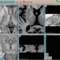
Stereotactic surgery in its early years mainly relied on ventriculocisternography to localize internal structures for lesioning, but by the late 1970s, target localization in stereotactic surgery was mainly done by CT imaging. A localizing plate with vertical fiducial posts filled with radio-opaque material was fixed to the base of the stereotactic frame. These posts essentially formed a picket fence around the perimeter of the stereotactic frame while the patient was being imaged. The positions of the posts were read as the x–y coordinates in the CT images and used as reference points. The location of the target was then triangulated. By late 1970s, the accuracy of target localization was greatly improved through the innovation of Russell Brown who added a diagonal bar across the two picket posts to generate an N-localizer. This ingenious design simplified the conversion of the two-dimensional coordinates into three-dimensional reference points in the CT images (Fig. 3.3). The N-localizer allows the colocalization of the desired target in both CT and MRI images, and improves the accuracy of multimodal image fusion. In the 1980s the N-localizer was universally adapted and was incorporated into the design of the Leskell frame for stereotactic radiosurgery [10–12].

Fig. 3.3


The use of a N-localizer (a) to translate two-dimensional coordinates into three-dimensional reference points in the CT images (b). (c) The localization of the target can then be checked against a phantom base
Related posts:


Stay updated, free articles. Join our Telegram channel
Full access? Get Clinical Tree
