Tension Pneumothorax
Peter J. Noone
Katherine R. Birchard
CLINICAL HISTORY
26-year-old female presents with shortness of breath and left chest pain after bronchoscopy earlier the same day.
 FIGURE 78A |
FINDINGS
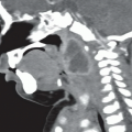
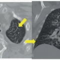
Figure 78A: Upright PA chest X-ray: Abnormal lucency in the left hemithorax, with partially collapsed left lung. Note visceral pleural edge (arrow) of left upper lobe, and mediastinal shift toward the right, and increased space between the left ribs compared to right. Axial contrast-enhanced chest CT: There is a large left pneumothorax (ptx), and the heart and mediastinum are shifted across midline. The left lower lobe is collapsed (star), and air fills remainder of hemithorax.
DIFFERENTIAL DIAGNOSIS
Pneumothorax, tension pneumothorax, collapsed right lung, pneumomediastinum, Poland syndrome.
DIAGNOSIS
Tension pneumothorax.
DISCUSSION
Clinical signs of a tension pneumothorax include tachypnea, hypotension, pleuritic chest pain, tracheal deviation, jugular vein distention, and unilateral absence of breath sounds. They occur when a perforation of the visceral pleura occurs with continuous air leak, but without chest wall injury. This causes accumulation of air in one hemithorax with subsequent increased pressure, compressing the lung and mediastinal structures to the contralateral side. Tension pneumothoraces often occur after trauma. Other risk factors include recent procedure such as central line placement, bronchoscopy, mechanical ventilation, and underlying lung disease. If a tension pneumothorax is strongly suspected, radiologic evidence is not necessary and the clinician should proceed with a needle thoracostomy at the second or third intercostal space at the midclavicular line to decompress the thorax followed by chest tube placement once the patient is stabilized. For patients in whom the diagnosis is less certain, chest radiograph is the initial study of choice. Findings suggesting a tension pneumothorax include hyperlucency in one hemithorax with increased intercostal space compared to the contralateral hemithorax, and tracheal, esophageal, and mediastinal deviation away from the pneumothorax.
Related posts:


Stay updated, free articles. Join our Telegram channel

Full access? Get Clinical Tree


