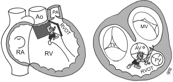
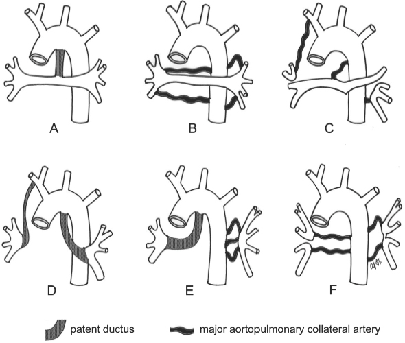
15 Tetralogy of Fallot and Related Conditions Fig. 15.1 Features of tetralogy of Fallot. Ao, ascending aorta; AV, aortic valve; MV, mitral valve; PA, main pulmonary artery; PV, pulmonary valve; RA, right atrium; RV, right ventricle; RVOT, right ventricular outflow tract. Fig. 15.2 In severe form, tetralogy is associated with pulmonary atresia. Pulmonary circulation may be supplied by a patent ductus arteriosus or major aortopulmonary collateral arteries. The right and left pulmonary arteries can be confluent (A–C) or nonconfluent (D–F). Not infrequently, branch pulmonary arteries in the mediastinum are not formed (E and F).
Definition and Classification
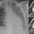
 Tetralogy of Fallot is composed of a constant set of the following four features, which result from anterior, superior, and leftward displacement of the outlet or infundibular septum relative to the rest of the septum (Fig. 15.1). The displaced outlet septum (door) encroaches on the right ventricular outflow tract (RVOT).
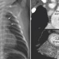
 In severe form, tetralogy is associated with pulmonary atresia. The pulmonary arterial anatomy varies and the blood flow to the pulmonary arteries is through either a patent ductus arteriosus or collateral arteries arising from the aorta or its branches (Fig. 15.2). The collateral arteries are of congenital origin and called major aortopulmonary collateral arteries (MAPCAs) in contrast to acquired collateral arteries such as dilated bronchial and intercostal arteries.
 Tetralogy of Fallot is rarely associated with absent pulmonary syndrome in which the RVOT is guarded by vestigial valve tissue causing free pulmonary regurgitation from in utero life. The proximal pulmonary arteries in the mediastinum and lung hila are aneurysmally dilated.
 A large ventricular septal defect can be associated with an obstructing aberrant muscle bundle within the right ventricle above the septal defect, causing a hemodynamic effect similar to that in tetralogy of Fallot. This condition is called two- or double-chambered right ventricle.
Pathophysiology
 Hemodynamics of tetralogy are dependent on the degree of pulmonary stenosis and pulmonary arterial anatomy.
 Right-to-left shunt through the ventricular septal defect
 The more severe the pulmonary stenosis, the greater the right-to-left shunt through the ventricular septal defect, allowing blood flow from the right ventricle into the overriding aorta and reducing pulmonary blood flow.
 Mild pulmonary stenosis with a large ventricular septal defect resembles an isolated ventricular septal defect. This mild form is termed an acyanotic or pink tetralogy of Fallot.
 Atretic pulmonary outflow tract may be considered a severe form of tetralogy of Fallot and may involve the pulmonary valve and variable portions of the pulmonary arteries. In severe forms, the branch pulmonary arteries are involved, and the continuity between right and left pulmonary arteries is interrupted (Fig. 15.2).
 In pulmonary atresia, pulmonary circulation may be supplied through a patent ductus arteriosus or systemic collateral arteries. In most cases, the patent ductus arteriosus and the MAPCAs do not coexist in supplying the same lung. Patent ductus arteriosus or MAPCAs are rarely seen in cases without pulmonary atresia. Morphology of pulmonary arterial tree and supply are the central features (Fig. 15.2).
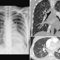
 Absent pulmonary valve variant is characterized by severe pulmonary regurgitation and severely dilated central pulmonary arteries, which cause significant compression of the main and segmental bronchi.
 Peripheral pulmonary arteries show arborization abnormalities and may compress more distal bronchi, leading to distal air trapping
 Central bronchi may have deficient or defective cartilage also leading to bronchomalacia.
 Tetralogy of Fallot is often associated with deletion of chromosome 22q11, and less commonly with the VAC-TERL association (vertebral abnormalities, anal atresia, cardiac abnormalities, tracheoesophageal fistula, esophageal atresia, renal agenesis, and limb defects) or trisomy 21.
Clinical Manifestations
Related posts:
Stay updated, free articles. Join our Telegram channel

Full access? Get Clinical Tree


