, Marilyn J. Siegel2, Tomasz Miszalski-Jamka3, 4 and Robert Pelberg1
(1)
The Christ Hospital Heart and Vascular Center of Greater Cincinnati, The Lindner Center for Research and Education, Cincinnati, OH, USA
(2)
Mallinckrodt Institute of Radiology, Washington University School of Medicine, St. Louis, Missouri, USA
(3)
Department of Clinical Radiology and Imaging Diagnostics, 4th Military Hospital, Wrocław, Poland
(4)
Center for Diagnosis Prevention and Telemedicine, John Paul II Hospital, Kraków, Poland
Abstract
The classic lesions of tetralogy of Fallot (TOF) include pulmonary stenosis, ventricular septal defect (VSD), overriding aorta, and right ventricular hypertrophy. The hemodynamically significant lesions are the VSD and pulmonary stenosis that results in right ventricular outflow tract (RVOT) obstruction. Originally, a two-stage repair was performed for repair of TOF. First pulmonary blood flow was increased with systemic-to-pulmonary artery shunts (classic and modified Blalock–Taussig shunt, Waterston shunt, or Potts shunt) followed by a complete repair when the child was older. Complications associated with this strategy include pulmonary hypertension and congestive heart failure from the augmented pulmonary blood flow and stenosis of the pulmonary artery at the anastomotic site.
The classic lesions of tetralogy of Fallot (TOF) include pulmonary stenosis, ventricular septal defect (VSD), overriding aorta, and right ventricular hypertrophy. The hemodynamically significant lesions are the VSD and pulmonary stenosis that results in right ventricular outflow tract (RVOT) obstruction. Originally, a two-stage repair was performed for repair of TOF. First pulmonary blood flow was increased with systemic-to-pulmonary artery shunts (classic and modified Blalock–Taussig shunt, Waterston shunt, or Potts shunt) followed by a complete repair when the child was older. Complications associated with this strategy include pulmonary hypertension and congestive heart failure from the augmented pulmonary blood flow and stenosis of the pulmonary artery at the anastomotic site.
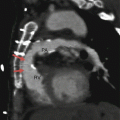
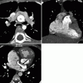
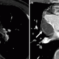
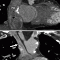
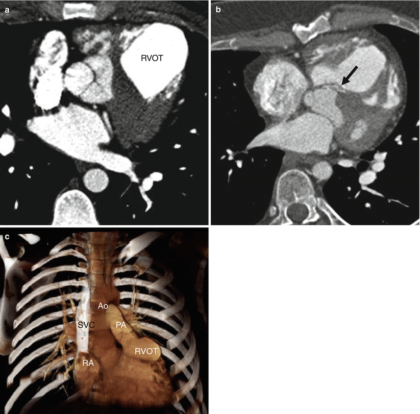
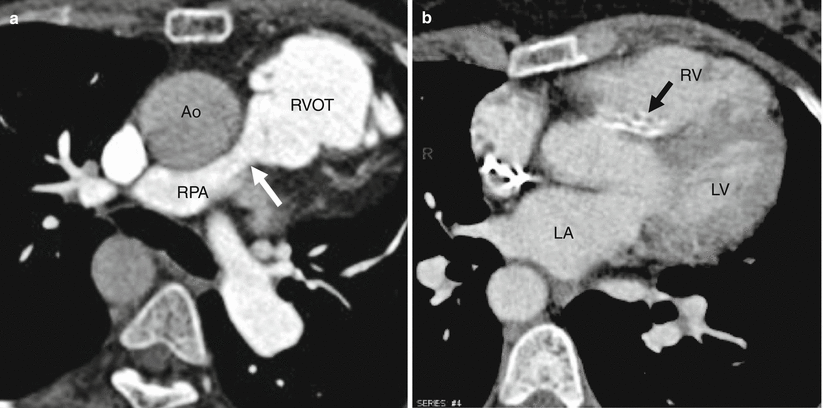
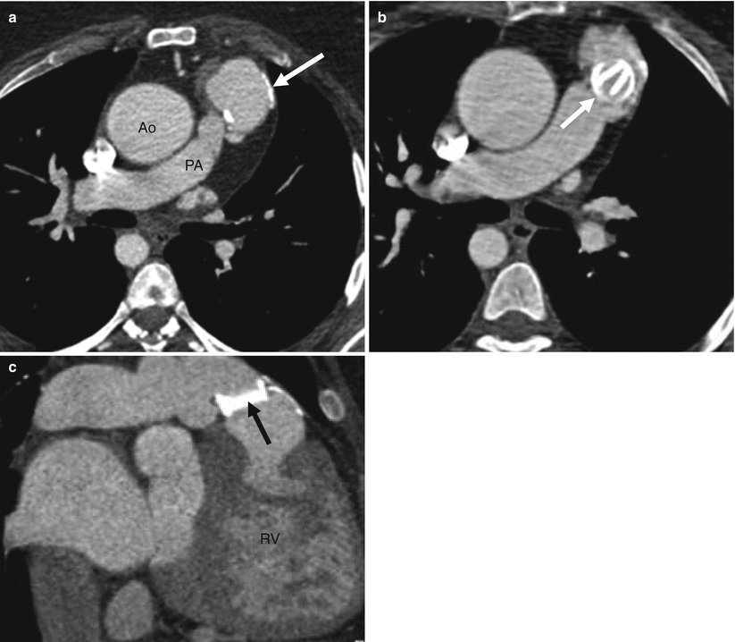
The current treatment for TOF with pulmonary stenosis is a primary single-stage repair in the first year of life, usually at 3–6 months of age. This obviates the need for multiple surgeries and prevents the associated complications. Definitive repair consists of closing the VSD and relieving the RVOT obstruction. The older surgical technique relieved the RVOT obstruction using a right ventricular free-wall incision with insertion of an infundibular patch graft or homograft to enlarge the outflow tract and also included a repair of the VSD with a GORE-TEX patch (Figs. 29.1 and 29.2). The pulmonary homograft replaced the pulmonary valve and proximal pulmonary artery. This repair is no longer performed due to the frequent complication of dilatation of the RVOT homograft leading to progressive pulmonary valve regurgitation requiring valve replacement and the development of right ventricular failure (Fig. 29.3).
Get Clinical Tree app for offline access

Fig. 29.1
Repaired tetralogy of Fallot, using a patch to close the ventricular septal defect and a homograft to enlarge the right ventricular outflow tract. Panel (a) is an axial scan showing a right ventricular outflow tract (RVOT) homograft. Notice mild dilatation of the graft. Panel (b) is an axial cut showing a high-density ventricular septal defect patch (arrow). Panel (c) is a coronal image showing the mildly dilated RVOT and proximal pulmonary artery (PA). SVC superior vena cava, PA pulmonary artery, Ao aorta

Fig. 29.2
Repaired tetralogy of Fallot in a 54-year-old woman who has mild pulmonic regurgitation and normal right ventricular function. Panel (a) is an axial scan showing enlargement of the right ventricular outflow tract (RVOT) homograft. Also note the narrowing at the junction of the right pulmonary artery (RPA) with the homograft (white arrow). Panel (b) an axial image showing calcification in the ventricular septal defect patch (arrow)

Fig. 29.3
Repaired tetralogy of Fallot complication by dilatation of the right ventricular outflow tract patch graft leading to wide open pulmonary valve regurgitation and mechanical valve replacement. Panel (a) is an axial image showing a calcified, dilated right ventricular outflow tract (arrow). Also note the right-sided aortic arch and dilatation of the ascending aorta (Ao) leading to aortic regurgitation. Panel (b


Stay updated, free articles. Join our Telegram channel

Full access? Get Clinical Tree


