Chapter Outline
Omental Torsion and Infarction
Hepatosplenic Vascular Disease
The term acute abdomen defines a clinical syndrome characterized by the sudden onset of severe abdominal pain requiring emergency medical or surgical treatment. A prompt and accurate diagnosis is essential to minimize morbidity and mortality. The differential diagnosis includes an enormous spectrum of infectious, inflammatory, obstructive, and neoplastic disorders ranging from benign self-limited diseases to conditions that require emergency surgery ( Fig. 124-1 ). In a review of approximately 30,000 patients with acute abdomen, Shah observed that 28% of patients had appendicitis, 9.7% had acute cholecystitis, 4.1% had obstruction of the small bowel, 4% had acute gynecologic disease, 2.9% had acute pancreatitis, 2.9% had acute renal colic, 2.5% had perforated peptic ulcer, and 1.5% had diverticulitis. For one third of patients, no cause could be determined.

The clinical diagnosis of acute abdomen can be challenging because results of physical examination, clinical presentation, and laboratory examination are often nonspecific and nondiagnostic. Sonography has developed a niche in evaluating the gallbladder in all patients and the appendix in children and pregnant women. Magnetic resonance (MR) is being used with increasing frequency in pregnant women. Multidetector computed tomography (MDCT), however, has become the premier technique for triage of most patients with acute abdomen. MDCT has earned this role because it can provide a global perspective of the gut, mesenteries, omenta, peritoneum, retroperitoneum, subperitoneum, and extraperitoneum uninhibited by the presence of bowel gas and fat. MDCT scanning allows thinner contiguous images to be obtained without increasing radiation exposure and without respiratory misregistration. Multiplanar reformatted images can be obtained with virtually isotropic data sets. The rapidity of scanning allows several acquisitions to be obtained during different phases of a single intravenous bolus of contrast material. The cross-sectional imaging features of the most common abdominal disorders causing the acute abdomen are discussed in this chapter.
Technical Considerations
A variety of MDCT protocols for preparation of the patient and scanning have been created to study the diversity of diseases that can cause acute abdomen. The selection of an imaging technique depends on the most likely diagnosis, clinical setting, and local expertise. The examination should be tailored to each patient. See Chapter 5 for a more complete discussion of abdominal CT protocols.
It is best to obtain a general survey examination that includes the entire abdomen and pelvis. Diagnostic errors will occur if the anatomic coverage is dictated solely by the vagaries of clinical diagnosis. Scans are obtained from the diaphragm to beneath the symphysis pubis. Coronal and sagittal reformatted images are helpful in establishing a diagnosis.
Intravenous administration of contrast material is helpful in the diagnosis of splanchnic venous thrombosis, bowel ischemia, aneurysms, and active arterial extravasation as well as solid parenchymal organ abnormalities. Inflammatory mural changes in appendicitis, cholecystitis, diverticulitis, Crohn’s disease, and infectious enterocolitis are also better depicted with vascular enhancement. Neoplasms, abscesses, and infarcts in the liver, spleen, and kidneys are well portrayed on contrast-enhanced scans. Intravenously administered iodinated contrast material carries the risk of nephrotoxicity and potential reaction to the agent, and it may obscure renal and ureteral stones. However, in most patients, the information provided justifies the risk and extra expense. Between 125 and 150 mL of 60% iodinated contrast material should be injected intravenously at a rate of at least 3 mL/s. Scans are obtained during the portal venous phase with a 60- to 70-second delay. Arterial phase imaging (40-second delay) is useful in patients with suspected hemorrhage, bowel ischemia, and arterial thrombosis. Delayed scans through the kidneys and pelvis can reveal pyelonephritis, renal masses, and bladder disease that might have been overlooked during earlier phases.
When bowel obstruction, intestinal ischemia or infarction, ileus, intestinal infection, or inflammation is suspected, the intrinsic fluid in the gut often serves as an excellent gastrointestinal luminal contrast agent. Positive contrast agents may lead to algorithm undershoot or overshoot and may interfere with assessment of bowel enhancement and viability. No oral contrast agent, water, or a low-contrast agent such as VoLumen may be given in these cases. For suspected kidney or ureteric stones and ruptured abdominal aortic aneurysms, scans without oral or intravenous administration of contrast material should first be obtained.
For patients with nonspecific symptoms and signs, we prefer to give 800 to 1000 mL of a 2% solution of oral, diluted, water-soluble contrast material at least 1 hour before scanning. Oral contrast material is administered primarily to differentiate bowel loops from abdominal and pelvic masses and abscesses. Oral contrast material may obscure the diagnosis of bowel hemorrhage or ischemia and limit the detection of ureteral stones, appendicoliths, and bile duct stones. Practical difficulties of using oral contrast material include the time it takes to opacify the gut, the randomness of contrast opacification, and the inability of sick patients to consume and to retain sufficient quantities of the agent.
The use of rectal contrast material is advocated by some investigators to optimize the detection of appendicitis, diverticulitis, and epiploic appendagitis. With the patient in the left decubitus position, 400 to 600 mL of a 3% solution of water-soluble contrast agent is administered by gravity through a soft rubber rectal catheter without use of a balloon. The patient is then turned to the supine position for scanning.
An alternative approach to the patient with acute abdomen is to perform CT without oral, intravenous, or rectal administration of contrast media. This technique is fast, is virtually risk free, and causes no patient discomfort. However, these scans are the most difficult to interpret, particularly in patients with little abdominal or pelvic fat.
Appendicitis
Acute appendicitis ( Fig. 124-2 ) is the most common abdominal surgical emergency, affecting approximately 250,000 people annually in the United States. Although the correct diagnosis can be made in most patients on the basis of the history, physical examination findings, and laboratory test results, the diagnosis is uncertain in 20% to 33% of patients who present with atypical symptoms. The diagnosis is most difficult for infants, young children, elderly patients, and women of reproductive age. In the past, an average negative laparotomy rate of 20% was acceptable. The widespread use of MDCT for patients with suspected appendicitis positively affects patient outcomes and increases the number of laparotomies with positive results. The surgical misdiagnosis rate has been reduced from 20% to 40% without imaging to a current rate of approximately 5% to 10%.

The MDCT, ultrasound, and MR findings of acute appendicitis reflect the extent and severity of inflammation ( Fig. 124-3 ). In mild disease, the appendix appears as a slightly distended (6-15 mm in diameter), fluid-filled structure that shows circumferential symmetric mural thickening ( Fig. 124-4 ). Sometimes, only the tip is inflamed (so-called tip appendicitis). On sonographic examination, periappendiceal inflammation may be encountered, and pain over the appendix may be elicited. The inflamed appendix is often hypervascular on color Doppler ultrasound.


Homogeneous, dense contrast enhancement of the wall is typical on MDCT, but a target sign may be seen on axial images (see Fig. 124-4 ). Periappendiceal inflammation is manifested as slight haziness of the mesoappendix fat. A calcified appendicolith is more reliably revealed on CT than on plain radiography. When it is present, the appendicolith is less well visualized than on CT or ultrasound. With disease progression and perforation, the appendix becomes fragmented, destroyed, and replaced by a phlegmon or abscess ( Fig. 124-5 ). Associated mural thickening of the adjacent distal ileum and cecum may also occur. In patients with these symptoms, the specific diagnosis of appendicitis can be made if an appendicolith is seen in the abscess or phlegmon. If not, the diagnosis of appendicitis can only be suggested within a differential diagnosis that includes cecal diverticulitis, ileal diverticulitis, Meckel’s diverticulitis, perforated neoplasm (cecal, appendiceal, or ileal), and Crohn’s disease with abscess formation.

MR ( Fig. 124-6 ) is becoming increasingly used in pregnant patients with high clinical suspicion of appendicitis. The MR features are similar to those seen on CT. The appendix is distended to a caliber greater than 6 to 7 mm in diameter with surrounding inflammatory change in the mesoappendix. See Chapter 56 for a more complete discussion of appendicitis.

Diverticulitis
Diverticulitis occurs in 10% to 25% of patients with known diverticulosis. It results from a microperforation or, much less commonly, a macroperforation of a diverticulum into the rich pericolic fat in the subperitoneal spaces surrounding the colon. These patients typically present with left lower quadrant pain, fever, and leukocytosis. Clinical misdiagnosis rates range from 34% to 67%. The role of CT for these patients is to confirm the diagnosis, to establish the presence of complications (e.g., abscess), to provide a road map for percutaneous or surgical therapy, and to suggest alternative diagnoses for patients in whom diverticulitis has been excluded.
The CT hallmark of diverticulitis is inflammatory change in the pericolic fat, which is observed in 98% of patients ( Fig. 124-7 ). Minimal haziness of adjacent fat occurs in mild cases. Small fluid collections, fine linear strands, and extraluminal gas bubbles may also occur. In more severe cases, phlegmon or frank abscess formation can occur. Diverticula are evident in more than 80% of patients, and symmetric mural thickening of more than 4 mm is seen in about 70% of patients. Other typical features include engorgement of the vasa recta and the presence of fluid in the inferior portion of the combined interfascial plane.

In some patients, contrast material collects in an arrowhead shape adjacent to the inflamed colonic diverticulum (i.e., arrowhead sign of diverticulitis). The offending inflamed diverticulum may appear as a rounded paracolic outpouching centered in the paracolic inflammation with soft tissue calcium, barium, or air attenuation.
A perforated carcinoma is the major differential diagnostic consideration for patients with sigmoid diverticulitis. Although the colon wall is usually less than 1 cm thick in acute diverticulitis, in patients with severe muscle hypertrophy, the wall may be 2 to 3 cm thick, simulating carcinoma. CT findings favoring the diagnosis of acute diverticulitis include a tethered or saw-toothed luminal configuration, the presence of fluid in the combined interfascial plane, and engorged vasa recta. An abrupt zone of transition with normal bowel, enlarged local lymph nodes, and mural thickness greater than 1.5 cm favors carcinoma.
Complications of acute diverticulitis include abscess formation ( Fig. 124-8 ), obstruction of the large and small bowel, secondary inflammation of the appendix, fistula, sinus tracks, and frank intraperitoneal perforation. Right-sided diverticulitis is usually difficult to diagnose clinically. Compared with patients with appendicitis, individuals with right-sided diverticulitis have a more protracted history, milder pain, and a higher point of maximum tenderness, which may clinically simulate acute cholecystitis. A palpable mass is present in up to one third of patients and can mimic an appendiceal or cecal tumor.

The MDCT findings of right-sided diverticulitis consist of focal pericolic inflammatory change, slight mural thickening, and visualization of diverticulum as an outpouching of the right colon at the level of maximum wall thickness. The offending diverticulum contains gas, fluid, contrast material, or calcified material. The normal appendix should be seen. If the appendix is not visualized, appendicitis, epiploic appendagitis, typhlitis, or perforated cecal carcinoma must be considered in the differential diagnosis. See Chapter 55 for a more complete discussion of diverticulitis.
Bowel Obstruction
Obstruction of the small intestine and colon accounts for approximately 20% of acute abdominal surgical conditions. MDCT has replaced conventional contrast studies because it can more reliably answer several questions. Is obstruction present? What is the level of obstruction? What is the cause of obstruction? What is the severity of obstruction? Is the obstruction simple or a closed loop? Is strangulation or ischemia present?
It is important to differentiate between simple and closed-loop obstruction ( Fig. 124-9 ) because the simple obstruction can be treated conservatively, whereas closed-loop obstruction requires prompt surgical intervention. For patients with bowel obstruction, scans are best obtained without oral contrast material because intraluminal fluid and gas serve as natural contrast agents. Intravenous contrast material is important in assessing intestinal perfusion and ischemia and in delineating the size, configuration, and patency of the mesenteric vessels. If oral contrast material is given, a delayed plain abdominal radiograph obtained several hours later can determine if the contrast material has passed into the colon.

The CT hallmark of bowel obstruction is the delineation of a transition zone between dilated and decompressed bowel. Careful inspection of the transition zone and luminal contents often reveals the underlying causes of obstruction. CT is most helpful in patients with internal and external hernias, neoplasms, gallstone ileus, various forms of enteroenteric intussusception, and afferent loop obstruction after a Billroth II operation. If no mass, hernia, intussusception, abscess, or inflammatory thickening is present, adhesion is the most likely diagnosis. The typical adhesion has a beaklike narrowing, and the affected gut may be difficult to view, depending on the orientation of the loop relative to the axial plane. Use of the scroll or leaf function and multiplanar reformations in the coronal and sagittal planes can help establish the correct diagnosis.
The anterior abdominal wall should be carefully inspected to search for prior surgical scars. The small bowel feces sign is often seen just proximal to the obstruction. If positive oral contrast material is present, it becomes progressively more dilute as it approaches the level of obstruction. Also the degree of small bowel dilation is largest closest to the level of obstruction.
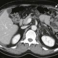
An incarcerated or closed-loop obstruction is manifested as a loop-shaped, fluid-filled structure causing proximal segments to dilate with gas and fluid. The mesenteric vessels have a radial distribution ( Fig. 124-10 ) because they become stretched and converge toward the U- or C-shaped loop. Two adjacent and collapsed round, oval, or triangular segments typically represent the afferent and efferent entry points of the torsion site. The mesenteric vasculature may have an unusual course. When ischemia develops, the bowel wall may thicken and have a target appearance caused by submucosal edema. Enhancement of the involved bowel wall may be poor or delayed. Fluid and hemorrhage may collect in the mesentery, bowel wall, and lumen of the involved segment. The mesentery becomes hazy in appearance, and ascites may develop.

In patients with high-grade obstruction of the small bowel, CT has a reported sensitivity of 90% to 99%. CT is less accurate in patients with low-grade obstruction. Obstruction of the large bowel ( Fig. 124-11 ) can also be demonstrated by MDCT. See Chapters 46 and 62 for a more complete discussion of bowel obstruction.

Acute Cholecystitis
Acute cholecystitis results from obstruction of the gallbladder and its attendant mural inflammation associated with infection and sometimes necrosis. Most cases are caused by obstructing gallstones in the gallbladder neck or cystic duct. Because acute cholecystitis develops in only 20% of patients with gallstones, many patients with gallstones with right upper quadrant pain have other pathologic conditions responsible for their symptoms.
Only 20% to 30% of patients with right upper quadrant pain have acute cholecystitis. The primary sonographic diagnostic criterion is the sonographic Murphy sign associated with gallstones. Secondary signs of acute cholecystitis include mural thickening (>3 mm) and stratification, a distended or hydropic gallbladder with loss of the normal tapered neck and development of an elliptical or rounded shape, and pericholecystic fluid ( Fig. 124-12 ).

Although sonography is the preferred method for diagnosis of acute cholecystitis, CT is frequently the initial examination because the diagnosis is unclear. The most sensitive CT findings of acute cholecystitis are mural thickening greater than 3 mm (in the setting of a distended gallbladder) and enhancement of the inflamed wall ( Fig. 124-13A ). Transient, focally increased attenuation of the liver may develop adjacent to the inflamed gallbladder, resulting from hepatic artery hyperemia and early venous drainage ( Fig. 124-13B ). Less specific signs include pericholecystic fluid, haziness of the pericholecystic fat, and increased attenuation of the gallbladder bile. CT can also depict complications of acute cholecystitis, including perforation and gangrene. Intramural or intraluminal gas is present in emphysematous cholecystitis. See Chapter 77 for a complete discussion of acute cholecystitis.

Choledocholithiasis
Patients with choledocholithiasis typically present with acute right upper quadrant pain, fever, jaundice, and pancreatitis. Thin-collimation scans are needed to optimize the detection of stones on MDCT. A high-density nidus may be visualized in the duct, or alternating low- and high-density rings of mixed cholesterol-calcium stones may be seen. Biliary dilation may be evident proximally. MDCT has a sensitivity of 88%, specificity of 97%, and accuracy of 94% in the detection of choledocholithiasis; however, positive intraluminal and intravascular contrast agents can obscure the detection of peripherally calcified stones. MR and MR cholangiopancreatography are the premier means of establishing the diagnosis of choledocholithiasis.
Peptic Ulcer Disease
Patients with peptic ulcer disease often present with nonlocalizing signs and symptoms indistinguishable from those of acute pancreatitis or cholecystitis, and MDCT is normally the first examination ordered. The most common MDCT result is focal mural thickening, which is a nonspecific finding. On occasion, an active ulcer or perforation ( Fig. 124-14 ) is identified, accompanied by inflammatory change of the adjacent fat, mesenteries, and omenta.