Chapter 1
The cross-sectional anatomy of the liver and normal variations
Ersan Altun1, Mohamed El-Azzazi1,2,3,4, and Richard C. Semelka1
1The University of North Carolina at Chapel Hill, Department of Radiology, Chapel Hill, NC, USA
2University of Dammam, Department of Radiology, Dammam, Saudi Arabia
3King Fahd Hospital of the University, Department of Radiology, Khobar, Saudi Arabia
4University of Al Azhar, Department of Radiology, Cairo, Egypt
Knowledge of cross-sectional anatomy of the liver is essential for the determination of localization of disease processes and for their management. To have a good knowledge and sense of the cross-sectional anatomy of the liver on computerized tomography (CT) and magnetic resonance imaging (MRI) studies, the segmental anatomy of the hepatic parenchyma and the anatomy of hepatic fissures, hepatic vessels, and bile ducts should be understood.
Liver anatomy can be described using two different approaches, including morphological anatomy and functional anatomy (1).
Morphological anatomy of the liver describes the liver anatomy depending on external appearance of the liver (1). Four lobes of the liver including the right, left, caudate and quadrate can be identified on the basis of the fissures of the liver surface (1). Morphological anatomy is not sufficient for the needs of modern radiology, hepatology, and hepatobiliary surgery.
Functional anatomy of the liver describes the functional segments of the liver on the basis of the anatomy of hepatic vessels and bile ducts (1). Functional anatomy is necessary to meet the needs of modern radiology, hepatology, and hepatobiliary surgery. Functional anatomy of the liver has been described by a number of different nomenclature systems for the determination of anatomic segments of the liver. A single, universally accepted classification system for the functional segmental anatomy of the liver does not exist. The Goldsmith and Woodburne system (1957), the Couinaud system (1957) and the Bismuth system (1982) are the most commonly used nomenclaturel systems (1).
Functional anatomy
The segments of the liver
The Bismuth system, which is a modified version of the Couinaud system, is the most commonly used anatomic nomenclature system, particularly in the United States. This hepatic segmental nomenclature system meets the needs of modern surgical techniques (Table 1.1) (1–5) and allows hepatobiliary surgeons, hepatologists, and radiologists to use a common nomenclature that meets their needs and enables them to understand each other.
Table 1.1 Description of the liver segments according to the three most commonly used nomenclature systems.
| Part | Nomenclature system | |||||
| N. Goldsmith and R. Woodburne (1957) | C. Couinaud (1957) | H. Bismuth (1982) | ||||
| Segment | Subsegment | Sector | Segment | Sector | Segment | |
| Dorsal | Caudate L. | Caudate L. | I | Caudate L. | I | |
| Left | Lateral | Superior | Lateral | II | Posterior | II |
| Inferior | Paramedian | III | Anterior | III | ||
| Left | Medial | Superior | IV | IVa | ||
| Inferior | IVb | |||||
| Right | Anterior | Inferior | Paramedian | V | Anteromedial | V |
| Superior | VIII | VIII | ||||
| Right | Posterior | Inferior | Lateral | VI | Posterolateral | VI |
| Superior | VII | VII | ||||
The three vertical planes (scissurae) hosting the hepatic veins, and a transverse plane passing through the right and left portal vein branches are used to describe the segments of the liver (1, 5).
The three vertical scissurae hosting the hepatic veins divide the liver into four sectors and a transverse plane passing through the right and left portal vein branches divides these sectors into the eight segments, which are numbered clockwise on the frontal view. These segments can be described in a straightforward approach by combining the definitions of two systems including the Bismuth, and Goldsmith and Woodburne systems (Table 1.1). These liver segments, including the caudate lobe, can be described on the basis of this approach as follows: caudate lobe (I), left lateral superior (II), left lateral inferior (III), left medial superior (IVa), left medial inferior (IVb), right anterior inferior (V), right posterior inferior (VI), right posterior superior (VII), and right anterior superior (VIII) (Figures 1.1 and 1.2).
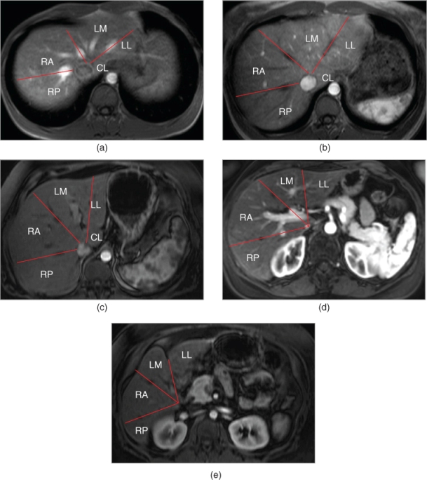
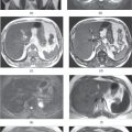
Figure 1.1 Segments of the Liver. T1-weighted axial hepatic venous (a) and hepatic arterial dominant (b–e) phase 3D-GE images acquired at different levels demonstrate the segments of liver, which are determined based on the distribution of diagonal planes (lines) hosting hepatic veins according to Goldsmith and Woodburne classification. RP, Right lobe posterior segment; RA, Right lobe anterior segment; LM, Left lobe medial segment; LL, Left lobe lateral segment; CL, Caudate lobe.
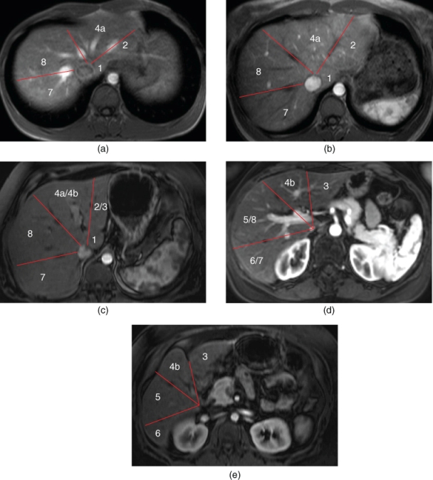
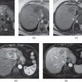
Figure 1.2 Segments of the Liver. T1-weighted axial hepatic venous (a) and hepatic arterial dominant (b–e) phase 3D-GE images acquired at different levels demonstrate the segments of liver, which are determined based on the distribution of diagonal planes hosting hepatic veins (lines) and transverse planes hosting portal veins according to Bismuth classification. 1: Caudate lobe. 2: Left Lateral inferior segment. 3: Left lateral superior segment. 4a: Left medial superior segment. 4b: Left medial inferior segment. 5: Right anterior inferior segment. 6: Right posterior inferior segment. 7: Right posterior superior segment. 8: Right anterior superior segment.
In the Bismuth system, each segment has an independent vascular supply, including arterial, portal, and venous supplies, as well as independent lymphatic and biliary drainage (1–5).
The caudate lobe has been described as a separate sector in the Bismuth system (1, 5). The caudate lob or segment I is located posteriorly, and positioned between the fissure for ligamentum venosum, the inferior vena cava (IVC), and porta hepatis (Figure 1.2) (1, 5). It is anatomically different from other segments as it may often have direct connections to the IVC through hepatic veins, which are different from the main hepatic veins (1, 5). The caudate lobe may also be supplied by both branches of the right and left hepatic arteries, and both branches of right and left portal veins (1, 5). Because of its different blood supply, the caudate lobe may be spared and/or may be hypertrophied to compensate for the loss of normal liver parenchyma in some liver disorders such as the Budd–Chiari syndrome or cirrhosis.
The corresponding branches of the hepatic arteries, portal veins, and tributaries of the bile ducts are intra-segmental and serve the corresponding segments of the liver by traveling together, while the hepatic veins run independently and are located inter-segmental (1, 5). The hepatic arteries, hepatic veins, portal veins, and bile ducts demonstrate frequent variations which may affect surgical procedures in liver transplantations and liver resections.
Normal variations of the liver segments
Related posts:

Stay updated, free articles. Join our Telegram channel

Full access? Get Clinical Tree


