The Detailed First Trimester Ultrasound Examination


The Detailed First Trimester Ultrasound Examination


INTRODUCTION

The role of the ultrasound examination in the first trimester has changed over the last 30 years with the introduction of nuchal translucency (NT) screening and with significant improvements in ultrasound technology. Ultrasound in early gestation, ranging from 6 to 16 weeks, was primarily performed to confirm cardiac activity, location of gestational sac, pregnancy dating, number of fetuses, and to assess the adnexal regions. In addition, ultrasound was used to guide invasive procedures such as chorionic villus sampling and amniocentesis. With the widespread use of the first trimester NT screening, the assessment of fetal anatomy became part of the early gestation ultrasound and many fetal malformations that were detected in the second and third trimesters of pregnancy are now detected in the first trimester. Major fetal anomalies such as fetal hydrops, anencephaly, body stalk anomaly, large anterior abdominal wall defects, megacystis, and others (see Table 5.1) are now almost universally detected in the first trimester.1 With accumulating knowledge and expanding expertise, the approach to the first trimester ultrasound has changed over time. The current goal of the first trimester ultrasound includes an element of fetal anatomic assessment and in experts’ hands, detailed evaluation of fetal anatomy is achievable and detection of several major fetal malformations is now possible with consistency. Advantages of the fetal anatomic survey in the first trimester include the ability to image the fetus in its entirety in one view, lack of bone ossification which obstructs view later in gestation, increased fetal mobility, which allows imaging from many different angles, and the availability of high-resolution transvaginal ultrasound, which brings the ultrasound transducer in proximity to fetal organs. Challenges of the first trimester anatomic survey however include the need to combine the abdominal and transvaginal approach in some cases, the small size of fetal organs, and the lack of some sonographic markers of fetal abnormalities that are commonly seen in the second trimester of pregnancy. In our experience, the performance of the fetal anatomic survey in the first trimester is enhanced if a systematic approach is employed.


Table 5.1 • Major Fetal Abnormalities That can Potentially be Easily Identified on First Trimester Ultrasound


Early hydrops


Anencephaly


Alobar holoprosencephaly


Body stalk anomaly


Ectopia cordis


Large omphalocele


Large gastroschisis


Megacystis


Molar placenta


In this chapter, we report on our systematic approach to the detailed fetal anatomic survey in the first trimester, defined at 11 to 14 weeks of gestation. We coined the term detailed to reflect on the comprehensive nature of this approach to fetal anatomy in the first trimester. This systematic approach is modeled along the “morphology/anatomy” ultrasound examination in the second trimester. It is important to emphasize that the performance of the detailed first trimester ultrasound examination requires substantial operator expertise in obstetric sonography, high-resolution ultrasound equipment, and knowledge of the current literature on this subject. Optimizing the first trimester ultrasound examination as described in Chapter 3 of this book, along with the use of the transvaginal approach with color Doppler and three-dimensional (3D) ultrasound when clinically indicated, will enhance its accuracy. In Chapter 1, we listed existing national and international guidelines for the performance of the first trimester ultrasound examination. The systematic approach that is proposed in this chapter expands on existing guidelines and is geared toward a detailed evaluation of fetal anatomy in early gestation. We have developed this approach to the detailed first trimester ultrasound over several years and have found it to be effective in screening for
fetal malformations in early gestation. Undoubtedly, as new information comes about and with technological advances in ultrasound imaging, the approach to the detailed first trimester ultrasound examination will evolve over time.


DETECTION OF FETAL MALFORMATIONS IN THE FIRST TRIMESTER

Over the past 25 years, several studies reported on the feasibility of ultrasound for the detection of fetal malformations in early gestation.2, 3, 4, 5, 6, 7, 8, 9, 10, 11, 12, 13, 14 Studies on this subject varied with some reporting high detection rates of fetal anomaly by ultrasound in early gestation performed in specialized centers with significant expertise.9,12,15, 16, 17, 18 Only few studies reported on the early gestation detection of fetal malformations in large screening populations with ultrasound examinations performed by several examiners with various level of expertise.1 Furthermore, the gestational age window varied between studies, with some reporting detection rates up to 16 weeks of gestation as part of the “early gestation ultrasound” whereas others have limited the ultrasound examination to the 11 to 14 weeks of gestation. Of particular interest is the comprehensive study of Syngelaki et al.,1 which reported on the detection of fetal anomalies during the 11- to 13-week scan in a population of 44,859 patients after excluding 332 detected aneuploidies.1 The authors classified the type of first trimester fetal malformations into four main groups: always detectable, occasionally detectable, rarely detectable and non-detectable.1 Table 5.2 summarizes the results of this study.1


Table 5.2 • Diagnosis of Fetal Anomalies at 11 to 13 Weeks Scan, after Excluding Detected Aneuploidies (N = 44,859)


Fetal Abnormality


Diagnosis at 11-13 Weeks in Relation to all Presenta


Neural Tube, Brain, Faceb


Acrania/iniencephaly


29/29 (100%)


Open spina bifida


3/21 (14.3%)


Ventriculomegaly


1/11 (9.1%)


Alobar holoprosencephaly


2/2 (100%)


Facial clefts


1/20 (5%)


Lungs, Heartc


Diaphragmatic hernia


4/8 (50%)


Cardiac anomalies (all)


28/106 (26.4%)


Abdomen, Renald


Omphalocele


60/60 (100%)


Gastroschisis


19/19 (100%)


Megacystis


29/29 (100%)


Infantile polycystic kidneys


2/6 (33.3%)


Skeletone


Lethal skeletal dysplasia


3/6 (50%)


Short long bones unilateral


2/4 (50%)


Absent hand/or foot


7/9 (77.8%)


Polydactyly


12/20 (60%)


Others and Multiple Anomalies


Body stalk anomaly


5/5 (100%)


Cloacal defect


1/1 (100%)


Multiple anomalies


8/8 (100%)


a In cases not diagnosed, absolute number are reported.

b None diagnosed at 11-13 weeks: Hemivertebra (1), microcephaly (1), craniosynostosis (1), agenesis corpus callosum (10), semilobar holoprosencephaly (1), cerebellar hypoplasia (1), vermian agenesis (1), nasopharyngeal teratoma (1), retrognathia (1).

c None diagnosed at 11-13 weeks: Cystic adenomatoid malformation (4), extralobar sequestration (2), isolated ventricular septal defect (10), cardiac tumors (4).

d None diagnosed at 11-13 weeks: Bladder exstrophy (1), duodenal atresia (2), bowel obstruction (1), renal agenesis unilateral (6), renal agenesis bilateral (1), renal agenesis and multicystic (3), hydronephrosis unilateral and bilateral (11), multicystic unilateral and bilateral (17), duplex kidneys (12).

e None diagnosed at 11-13 weeks: Arthrogryposis (1), talipes unilateral and bilateral (38), ectrodactyly (1).


Modified from Syngelaki A, Chelemen T, Dagklis T, et al. Challenges in the diagnosis of fetal non-chromosomal abnormalities at 11-13 weeks. Prenat Diagn. 2011;31:90-102; copyright John Wiley & Sons, Ltd., with permission.


In our experience, there are four main pathways that result in the prenatal diagnosis of fetal malformations in the first trimester:


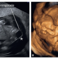
  • Major Malformation, Clearly Visible: The malformation is easily recognized during a routine first trimester ultrasound or an ultrasound performed for NT measurement, even with limited skills of the examiner.1,13 Table 5.1 summarizes some of the major anomalies that are clearly visible in the first trimester.


  • Thickened Nuchal Translucency: Many fetal malformations have been reported in association with thickened NT in the presence or absence of chromosomal aneuploidy. When a thickened NT is encountered, invasive genetic testing is typically offered along with an ultrasound examination of the fetus. This approach has led to the first trimester diagnosis of complex cardiac, brain, skeletal, gastrointestinal, and genitourinary anomalies as presented in various chapters in this book. On occasions, the associated fetal malformation is not seen in the first trimester but rather detected in the second trimester or even after birth and the relationship with an increased NT is thus assumed. Table 9.3 in Chapter 9 summarizes fetal malformations that are known to be associated with thickened NT.


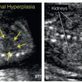
  • Pregnancies at High Risk for Fetal Malformations: When the pregnancy is at high risk for fetal anomaly due to a prior history of an affected child or due to a known inheritance pattern of a specific malformation, a detailed ultrasound in the first trimester can identify the fetal malformation. Examples include a pregnancy with prior spina bifida, an autosomal recessive inheritance pattern identified in a prior pregnancy, or an autosomal dominant inheritance pattern present in one of the parents. The presence of subtle findings in the first trimester ultrasound can be of significance in such cases such as the presence of abnormal intracranial translucency, polydactyly, echogenic kidneys, skeletal abnormalities, and cleft lip and palate, among others. Several of these subtle findings are discussed in detail in various chapters in this book.


  • Detailed First Trimester Ultrasound in Low-Risk Pregnancies: The detection of fetal malformations in the first trimester can also be the result of a detailed ultrasound examination that is routinely performed beyond the 11th week of gestation.11,13,19 With increasing skills and expertise in the detailed first trimester ultrasound, sonographers and sonologists may decide to apply this approach to all first trimester pregnancies beyond the 11th week of gestation for fetal anomaly screening. The detailed first trimester ultrasound will thus be an adjunct to the second trimester ultrasound examination. It is important to note however that several limitations currently exist to the detailed first trimester ultrasound examination and it is thus important to list these limitations before its introduction.


LIMITATIONS OF THE DETAILED FIRST TRIMESTER ULTRASOUND


Maternal Aspects

One of the main limitations of the detailed first trimester ultrasound examination is related to the accessibility of the relatively small gestational sac by ultrasound when maternal body habitus is increased, in the presence of prior abdominal surgery with scarring, and/or in the presence of large leiomyomas with posterior shadowing. In such conditions, the use of the transvaginal approach or a repeat ultrasound examination at 16 weeks of gestation either with a transabdominal high-resolution linear probe or with the transvaginal approach, if feasible, may provide sufficient access to assess fetal anatomy in detail. Occasionally, however, transient maternal contractions may trap the fetus in one area of the uterus and limit ultrasound accessibility. In our experience, rescanning the patient 15 to 30 minutes later provides for a better access, because in most cases the uterine contractions will have resolved (Fig. 5.1).


Indirect Signs of Fetal Malformations

Another limitation of the detailed first trimester ultrasound examination is the absence of classic, indirect signs of fetal malformations that are commonly seen in the second trimester.
For instance, unlike in the second trimester, bilateral renal agenesis is commonly associated with normal amniotic fluid volume in early gestation and open spina bifida does not typically display a lemon or banana sign in the first trimester as is very often seen in the second trimester of pregnancy. Other examples include the absence of hyperechogenicity or cystic changes in lung lesions in the first trimester and the lack of reliance on the abnormal cavum septi pellucidi in several central nervous system (CNS) lesions as commonly used in the second trimester ultrasound. Furthermore, fetal biometric changes and growth restriction that are commonly associated with fetal malformations do not manifest in early gestation and cannot be used as clues to the presence of associated malformations. It is important to note for all these reasons and others that the detailed first trimester ultrasound examination for fetal anatomy survey does not replace the traditional second trimester ultrasound but rather is complementary to it, especially in a high-risk pregnancy. Pregnant women should be informed of these limitations.


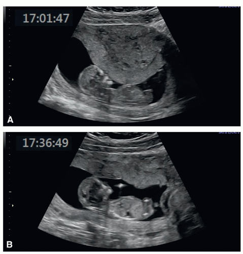
Figure 5.1: A: A transabdominal ultrasound examination in the first trimester showing a mid-uterine contraction, which is trapping the fetus in the midsection of the cavity. We were not able to complete the ultrasound examination despite an attempt by the transvaginal approach. The ultrasound examination was repeated 35 minutes later (B), which showed resolution of the contraction, optimization of imaging, and the fetus moving freely within the uterine cavity.


Time in Gestation of Development of Certain Malformations

It is important to note that a major limitation of the detailed first trimester ultrasound examination for fetal anatomy survey is that some ultrasound findings that are seen in early gestation may disappear upon follow-up into the second trimester of pregnancy. Examples include some cases of thickened NT, tricuspid regurgitation, cardiac ventricular disproportion, early fetal hydrops, and intraabdominal cystic lesions among others. On the other hand, some malformations that are traditionally visible in the second trimester, such as cystic lesions of lungs and kidneys, cardiac valvular stenosis, cortical brain abnormalities, cerebellar vermis dysgenesis, agenesis of corpus callosum, gastrointestinal atresias, and others are commonly associated with normal ultrasound findings in early gestation. It is therefore important for the sonographers and sonologists to be familiar with the natural course of congenital malformations and counsel patients regarding limitations of the first trimester ultrasound examination in that regard.


Safety Aspects

The detailed first trimester ultrasound examination is performed during a period of development and rapid growth of fetal organs. It is thus critical to minimize ultrasound exposure to the fetus, especially the use of pulsed Doppler, given its associated high energy. As discussed in detail in Chapter 2, the ALARA (as low as reasonably achievable) principle should always be followed and the operator should ensure that the thermal and mechanical indices levels always comply with safe practices. The risk of fetal exposure to ultrasound energy should always be balanced against the benefit of the ultrasound examination in early gestation. Refer to Chapter 2 of this book for a comprehensive discussion on ultrasound bioeffects and safety.


THE DETAILED FIRST TRIMESTER ULTRASOUND

In this section, we define our approach to the detailed first trimester ultrasound for fetal anatomy survey and describe the components of this ultrasound examination. As stated in the Introduction section of this chapter, the detailed first trimester ultrasound is performed at 11 to 14 weeks of gestation. Components of the detailed first trimester ultrasound examination include general overview and fetal biometry, comprehensive evaluation of fetal anatomy, and an assessment of the uterus and adnexal regions. This detailed first trimester ultrasound examination is not intended to replace the traditional second trimester ultrasound but rather to complement it, and in the majority of pregnancies to provide early reassurance of normalcy. The three components of the detailed first trimester ultrasound are described in the following sections of this chapter.


General Overview and Fetal Biometry

Initial aspects of the first trimester ultrasound include the confirmation of the location of the gestational sac within the endometrial cavity, the presence of cardiac activity, and the number of fetuses. This can be easily accomplished by the abdominal ultrasound, but on occasions may require the transvaginal approach. The position of the placenta in relation to the internal cervical os (Fig. 5.2) should be noted, keeping in mind that most placenta previas that are diagnosed in early gestation are of no clinical significance and will resolve upon follow-up ultrasound examination in the third trimester of pregnancy (see Chapter 15). In pregnancies with prior cesarean sections,
the scar is ideally assessed by transvaginal ultrasound.20 In these pregnancies the location of the gestational sac within the endometrial cavity should be noted and implantations in the lower uterine segment should raise suspicion for increased risk for placenta accreta21,22 (see Chapter 15). Furthermore, implantation of the gestational sac in the cesarean scar (cesarean scar implantation) is of significant importance given its association with placenta accreta and serious pregnancy complications.22,23 In the presence of twins or higher order multiple pregnancy, determining the chorionicity and amnionicity in the first trimester is of paramount importance. The presence and size of any significant subchorionic bleed (Fig. 5.3) should also be reported.


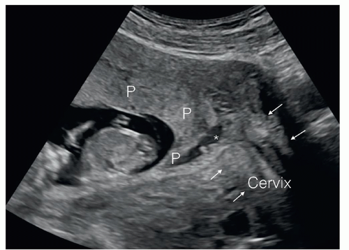
Figure 5.2: The position of the placenta (P) should be assessed in relation to the cervix (arrows) in the first trimester of pregnancy. This is often performed by the transabdominal ultrasound. Note that the placenta (P) is a previa in this pregnancy as it is shown to cover the internal cervical os (asterisk). The presence of placenta previa in the first trimester is of little clinical significance and should be followed up in the second trimester of pregnancy.


Figure 5.3: Subchorionic hematoma (arrows) in a pregnancy at 12 weeks of gestation in a patient presenting with vaginal bleeding. See text for details.


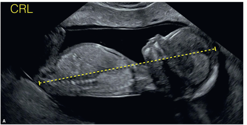
Figure 5.4: Fetal biometric measurements in the first trimester include crown-rump length (CRL) shown in A, biparietal diameter (BPD) and head circumference (HC) shown in B, abdominal circumference (AC) shown in C, and femur length (FL) shown in D.


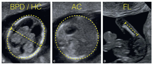
Figure 5.4: (continued)

Biometric measurements for pregnancy dating are an integral part of the first trimester ultrasound and include the measurement of the crown-rump length, the biparietal diameter, head circumference, abdominal circumference (AC), and femur length (FL) (Fig. 5.4). Any significant discrepancy in biometric measurements should alert for the possible presence of anatomic abnormalities or genetic malformations. First trimester fetal biometry and pregnancy dating are discussed in detail in Chapter 4. Table 5.3 lists the components of the general overview and fetal biometry of the detailed first trimester ultrasound.


Comprehensive Assessment of Fetal Anatomy

The comprehensive assessment of fetal anatomy is an important component of the detailed first trimester ultrasound. This approach to fetal anatomy in early gestation involves multiple sagittal, axial, and coronal planes of the fetus. Acquiring the technical skills required for the display of the corresponding anatomic planes and an in-depth knowledge of the current literature on this subject are prerequisites for the performance of the detailed first trimester ultrasound examination. In this section, we present our systematic approach to the assessment of fetal anatomy in the detailed first trimester ultrasound examination.


General Anatomic Assessment

The initial step of the fetal anatomy survey in the first trimester involves obtaining an anterior midsagittal plane of the fetus when technically feasible. This midsagittal plane allows for
a general anatomic assessment, given that the whole fetus is commonly included in this plane (Fig. 5.5). This midsagittal plane displays several important anatomic landmarks, which are listed in Table 5.4. In this midsagittal plane, the size and proportions of the fetal head, chest, and body are subjectively assessed and the following anatomic regions are recognized: fetal facial profile and midline intracranial structures, the anterior abdominal wall, the fetal stomach, and bladder. By slightly tilting the transducer from the midline to the left and right parasagittal planes, the arms and legs can be visualized. Many of the severe fetal malformations that can be detected in the first trimester (Table 5.1) will show abnormalities in the midsagittal
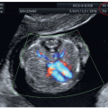
plane and an in-depth evaluation of fetal anatomic regions, as described in the following sections, will help to confirm the presence or absence of other fetal abnormalities. When clinically indicated, color and pulsed Doppler interrogation of the ductus venosus is also best assessed in this midsagittal plane.


Table 5.3 • General Overview and Fetal Biometry of the Detailed First Trimester Ultrasound


Location of gestational sac


Cardiac activity


Number of fetuses


Placental location in relation to cervix


Presence of subchorionic bleed


Fetal biometry

Only gold members can continue reading. Log In or Register to continue

Stay updated, free articles. Join our Telegram channel

Oct 14, 2019 | Posted by in ULTRASONOGRAPHY | Comments Off on The Detailed First Trimester Ultrasound Examination

Full access? Get Clinical Tree

Get Clinical Tree app for offline access