The Fetal Gastrointestinal System


The Fetal Gastrointestinal System


INTRODUCTION

Ultrasound examination of the fetal abdomen in the first trimester includes the assessment of abdominal organs from the diaphragm superiorly to the genitalia inferiorly. This ultrasound examination allows for the determination of fetal abdominal situs and for the anatomic evaluation of major organs in the gastrointestinal and genitourinary systems. This chapter focuses on the gastrointestinal tract, whereas the genitourinary system is discussed in the following chapter.


EMBRYOLOGY

The primitive gut is formed during the sixth menstrual week when the flat embryonic disc folds to form a tubular structure that incorporates the dorsal part of the yolk sac into the embryo (Fig. 12.1A-C). Ventral folding of the cranial, lateral, and caudal sections of the primitive gut forms the foregut, midgut, and hindgut, respectively (Fig. 12.2). In this process, the yolk sac remains connected to the midgut by the vitelline vessels (Fig 12.2). Three germ layers contribute to the formation of the gut, with the endoderm giving rise to the mucosal and submucosal surfaces; the mesoderm to the muscular, connective tissue and serosal surfaces; and the neural crest to the neurons and nerves of the submucosal and myenteric plexuses. The primitive gut is initially formed as a hollow tube, which is blocked by proliferating endoderm shortly after its formation. Recanalization occurs over the next 2 weeks by degeneration of tissue, and a hollow tube is formed again by the eighth menstrual week. Abnormalities of the recanalization process result in atresia, stenosis, or duplication of the gastrointestinal tract.


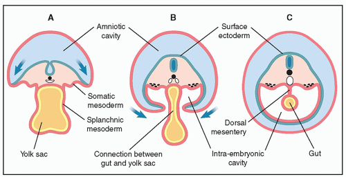
Figure 12.1: Axial views (A-C) of the developing embryo from the fourth week of gestation showing the formation of the primitive gut tube. Note the incorporation of part of the yolk sac into the embryo, shown in A and B and the primitive gut tube “gut” shown in C. See text for details.

The foregut, supplied by the celiac axis, gives rise to the trachea and respiratory tract (see Chapter 10), esophagus, stomach, liver, pancreas, upper duodenum, gall bladder, and bile ducts. The midgut, supplied by the superior mesenteric artery, gives rise to the lower duodenum, jejunum,
ileum, cecum, ascending colon, and proximal two-thirds of transverse colon. The hindgut, supplied by the inferior mesenteric artery, gives rise to the distal one-third of transverse colon, descending colon, sigmoid, rectum, and urogenital sinus. Because of lengthening of the gut and enlargement of upper abdominal organs, an intestinal loop from the midgut protrudes through the umbilical cord insertion into the abdomen at about the sixth week of embryogenesis (from fertilization). This intestinal loop returns to the intraabdominal cavity by about the 10th week of embryogenesis (from fertilization). Through the embryologic process, the midgut loop undergoes a series of three 90-degree counterclockwise rotations around the superior mesenteric artery.


Figure 12.2: Schematic drawing of a sagittal view of the embryo at approximately 5 to 6 menstrual weeks showing the formation of the primitive gastrointestinal tract (foregut, midgut, and hindgut) and the liver bud. Note the connection of the midgut to the vitelline duct. See text for details.


NORMAL SONOGRAPHIC ANATOMY

Sonographic visualization of the anatomy of the fetal abdomen is easily achieved in early gestation by the axial, sagittal, and coronal views of the fetus. We recommend a review of Chapter 5 on the systematic approach using the detailed first trimester ultrasound examination.


Axial Planes

The authors recommend the systematic evaluation of abdominal organs through three axial planes at the level of the upper abdomen (subdiaphragmatic—stomach) (Fig. 12.3), mid-abdomen (cord insertion) (Figs. 12.4 and 12.5), and the pelvis (bladder) (Fig. 12.6). In the upper abdominal axial plane, the fluid-filled anechoic stomach is imaged in the left upper abdomen and the slightly hypoechoic liver, as compared to the lungs, is seen to occupy the majority of the right abdomen (Fig. 12.3). The stomach is consistently seen at 12 weeks of gestation and beyond. This axial plane in the upper abdomen (Fig. 12.3) is important for the assessment of the abdominal situs (see later). In normal situs, the stomach occupies the left side of the abdomen, the liver and gall bladder occupy the right side of the abdomen, and the inferior vena cava (IVC) is seen anterior and to the right side of the descending aorta (Fig. 12.3B). The gall bladder is usually seen in about 50% of fetuses by the 13th week of gestation and practically in all fetuses by the 14th week of gestation.1 The mid-abdominal axial plane is important for the assessment of the cord insertion into the abdomen and the anterior abdominal wall (Fig. 12.4). In the mid-abdominal axial plane, the bowel is seen with a slightly hyperechoic sonographic appearance when compared to the liver (Fig. 12.4). Both kidneys can be seen in cross-section in the posterior aspect of the abdomen (Fig. 12.4A). It is important to note that physiologic bowel herniation is noted up until the 12th week of gestation (Fig. 12.5). Studies have shown that the midgut herniation should not exceed 7 mm in transverse measurements at any gestation and that the physiologic herniation is almost never seen at crown-rump length measurements exceeding 45 mm.2,3 The axial plane at the level of the pelvis reveals the bowel surrounding a small urinary bladder (Fig. 12.6). The two iliac crests can be seen in this plane in the posterior aspect of the pelvis (Fig. 12.6B). A slightly oblique plane of the pelvis in color Doppler demonstrates the two umbilical arteries surrounding the urinary bladder with an intact abdominal wall (Fig. 12.6B).


Figure 12.3: Axial planes at the level of the upper abdomen in two fetuses at 13 weeks of gestation. The fetus in A was examined transabdominally and the fetus in B transvaginally. Note the presence of fluid-filled stomachs (asterisks) in the upper left abdomen in A and B. Ribs (arrows) are visualized bilaterally along with the liver and inferior vena cava (IVC) in the right (R) abdomen. The descending aorta (DAo) is seen posterior and to the left of the IVC. Improved resolution is noted in fetus in B because of the transvaginal approach, thus allowing clear depiction of the IVC and DAo. L, left.


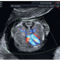
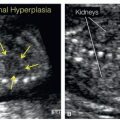
Figure 12.4: Axial plane of the middle abdomen in gray scale (A) and color Doppler (B) at the level of the umbilical cord attachment (arrow) in a fetus at 12 weeks of gestation. Note the presence of an intact anterior abdominal wall (arrow) and the fetal bowel appearing slightly more hyperechoic than surrounding tissue. Both kidneys (K) are seen in the posterior abdomen in A.


Sagittal Planes

In the sagittal and coronal planes of the fetus, the chest, abdomen, and pelvic organs are seen and are differentiated by their echogenicity. The lung and bowel are hyperechoic, the liver is hypoechoic, and the stomach and bladder are anechoic (Fig. 12.7). Lungs and liver are well separated by the concave-shaped diaphragm (Fig. 12.7). As in the second trimester, the parasagittal views do not exclude a diaphragmatic hernia. In the midsagittal view of the abdomen, the anterior abdominal wall with the umbilical cord insertion can be demonstrated (Fig. 12.7B) either on two-dimensional (2D) color Doppler or on three-dimensional (3D) ultrasound. This plane is ideally used in combination with color Doppler to visualize the course of the umbilical artery, vein, and ductus venosus (DV) (Fig. 12.8). The midsagittal plane of the abdomen is the most optimal plane for Doppler interrogation of the DV in early gestation (see Fig. 1.4).


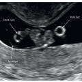
Figure 12.5: Axial views of the fetal abdomen in gray scale (A) and color Doppler (B) of a fetus at 10 weeks of gestation demonstrating the presence of a physiologic midgut herniation (arrow). In the corresponding 3D ultrasound in surface mode (C), the midgut herniation is shown as a bulge at the site of cord insertion into the abdomen (arrow).


Figure 12.6: Axial oblique plane of the lower abdomen at 13 weeks of gestation in gray scale (A) and color Doppler (B) demonstrating the fluid-filled urinary bladder (asterisk), surrounded by the left and right umbilical arteries (UA). This view is best visualized with color Doppler (B), which can also confirm the intact abdominal wall (arrow). Note the posterior location of the iliac crests in B.


Coronal Planes

A coronal view is rarely necessary in the first trimester, but it has been our experience that the coronal view is best suited to evaluate the position of the stomach when the diagnosis of diaphragmatic hernia is suspected (see Chapter 10). Transvaginal ultrasound examination of the abdomen in the first trimester provides high-resolution display of organs, which is helpful when abnormalities are suspected. It is important to note, however, that the fetal bowel appears more echogenic on transvaginal imaging, and differentiating normal bowel from hyperechogenic bowel because of pathologic conditions is difficult in early gestation. This is discussed later in this chapter.


Figure 12.7: Parasagittal plane in two fetuses at 13 (A) and 12 (B) weeks of gestation demonstrating the thorax and abdomen. The filled stomach (asterisk) is seen under the diaphragm (arrows). Fetus A is presenting in a dorso-posterior position and fetus B in a dorso-anterior position. Note the hyperechoic lungs and bowel, the hypoechoic liver, and anechoic stomach and bladder (not shown).


Figure 12.8: Midsagittal view of a fetus at 13 weeks of gestation in color Doppler demonstrating the cord arising from the abdomen (arrow) with the umbilical artery (UA) and vein (UV). Ao, descending aorta.


Figure 12.9: Schematic drawing (A) and corresponding 3D ultrasound image in surface mode of a fetus at 12 weeks of gestation. Note the normal insertion of the umbilical cord in the abdomen in A and B (arrows).


Three-Dimensional Ultrasound of the Fetal Abdomen

Similar to the use of 3D ultrasound in surface mode in the second and third trimester of pregnancy, 3D ultrasound in the first trimester provides additional information to the 2D ultrasound views.4 Surface mode is especially helpful for the demonstration of a normal and abnormal abdominal wall (Fig. 12.9), as illustrated in this chapter. For the assessment of the intraabdominal organs, 3D ultrasound can also be used in
multiplanar display, with reconstruction of planes for the specific evaluation of target anatomic regions displayed in tomographic view of axial (Fig. 12.10) or coronal (Fig. 12.11) planes. For more details on the use of 3D ultrasound in the first trimester, refer to Chapter 3 in this book and a recent book on the clinical use of 3D in prenatal medicine.4 Figures 12.5C and 12.9 show surface mode of the fetal anterior abdominal wall and Figures 12.10 and 12.11 show the use of multiplanar mode with plane reconstruction of axial and coronal views. In our experience, multiplanar mode can be of help especially in the transvaginal approach where transducer manipulation is limited (Figs. 12.10 and 12.11).


Figure 12.10: Tomographic axial views of the abdomen in a fetus at 12 weeks of gestation showing the upper, mid, and lower abdomen. Note the presence of the stomach (asterisk) and liver in the upper abdomen, kidneys (Kid.) and abdominal cord insertion (arrow) in the mid-abdomen, and the urinary bladder (Bl.) in the lower abdomen. L, left.


VENTRAL WALL DEFECTS

Defects of the abdominal wall are common anomalies in the fetus, and large defects are often detected in the first trimester.5 These anomalies include omphalocele, gastroschisis, Pentalogy of Cantrell, and body stalk anomaly (Table 12.1). Bladder exstrophy and cloacal exstrophy are often listed as abdominal wall defects, but are discussed in Chapter 13 as part of the urogenital anomalies.


Omphalocele


Ultrasound Findings

The physiologic midgut herniation (Fig. 12.5) is present between the 6th and 11th week of gestation and at crown-rump length of less than 45 mm.2 Therefore, the diagnosis of a small omphalocele cannot be performed reliably before the 12th week of gestation. The omphalocele is seen as a protrusion at the level of the cord insertion into the abdomen. The omphalocele-covering sac is seen as clear borders on ultrasound. Omphaloceles can be easily demonstrated on sagittal or axial views obtained at mid-abdomen (Figs. 12.12, 12.13, 12.14, 12.15 and 12.16). Figure 12.12 shows a schematic drawing of an omphalocele along with its corresponding 3D ultrasound in surface mode. In the first trimester, the omphalocele sac is either relatively small containing bowel loops (Fig. 12.13A) or large containing liver and bowel (Fig. 12.13B). Figure 12.14 shows parasagittal and axial views of a fetus with a large omphalocele, containing liver at 12 weeks of gestation, and Figure 12.15 shows a large omphalocele in two fetuses at 12 and 13 weeks of gestation containing bowel, liver, and stomach. The size of the omphalocele sac has an inverse relationship with chromosomal abnormalities. The presence of a small omphalocele in the first trimester with a thickened nuchal translucency should raise suspicion for the presence of associated fetal malformations and chromosomal aneuploidy (Figs. 12.13A and 12.16). Color Doppler helps in the demonstration of the umbilical cord attachment at the dome of the omphalocele, which can differentiate it from gastroschisis (Fig. 12.17). 3D ultrasound helps in the demonstration and documentation of the size of the omphalocele (Fig. 12.12). Transvaginal ultrasound provides detailed information of the omphalocele content and

additional anomalies of the heart, brain, kidneys, and spine. On occasion, the omphalocele can be as large or even larger than the abdominal circumference (Fig. 12.15). Follow-up of a first trimester isolated small omphalocele with a normal karyotype and nuchal translucency into the late second trimester is important because resolution of such cases has been documented in about 58% of fetuses.7 The presence of the liver in the omphalocele precludes resolution.


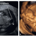
Figure 12.12: Schematic drawing (A) and corresponding 3D ultrasound image in surface mode of a fetus at 12 weeks of gestation with an omphalocele (arrows). Note in A the presence of an omphalocele sac covering the protruding intraabdominal organs (bowel, with or without liver), with the umbilical cord attached to the top of the omphalocele. The umbilical cord is not seen in B, as the lower extremities obscure it.


Figure 12.13: Midline sagittal plane in two fetuses with small (A) and large (B) omphalocele (arrows) at 12 weeks of gestation. In fetus A, the omphalocele is small and contains bowel only, whereas in fetus B, the omphalocele is relatively large and contains liver and bowel. Note the presence of an enlarged nuchal translucency (asterisk) in fetus A and workup revealed trisomy 18 in this fetus.


Figure 12.14: Parasagittal (A) and axial (B) view of a fetus at 12 weeks of gestation with a large omphalocele (arrows). Note the presence of liver and bowel within the omphalocele.


Figure 12.15: Axial view of the abdominal wall at the level of the cord insertion in two fetuses at 12 (A) and 13 (B) weeks of gestation. Note the presence of a large omphalocele (asterisks) with liver and bowel content in both fetuses. In fetus A the stomach is partly in the omphalocele, whereas in fetus B the stomach has completely protruded into the omphalocele. Sp, spine.


Figure 12.16: Axial view of the abdominal wall at the level of the cord insertion in two fetuses at 12 (A) and 13 (B) weeks of gestation. Note the presence of a small omphalocele (arrows) in fetus A and B, with only bowel content. Trisomy 18 was diagnosed in both fetuses.


Figure 12.17: Sagittal (A) and axial (B) planes of the mid-abdomen in color Doppler in a fetus at 12 weeks of gestation with trisomy 18. Note the presence of a small omphalocele (asterisk) in A and B and a thickened nuchal translucency (double headed arrow) in A. The use of color Doppler is helpful because it shows the umbilical cord arising from the top of the omphalocele in A (arrow) (compare with Fig. 12.12A) and a single umbilical artery in B (arrow).


Associated Malformations

Associated anomalies are common and are present in the majority of omphaloceles.6 Cardiac malformations are the most common associated structural abnormalities, and detailed cardiac imaging is thus recommended when an omphalocele is diagnosed in the first trimester6,8 (see Chapter 11). Chromosomal abnormalities, commonly trisomies 18, 13, and 21, are seen in about 50% of cases diagnosed in the first trimester.8 Omphaloceles associated with chromosomal abnormalities are typically small, with thickened nuchal translucency and other fetal structural abnormalities. Trisomy 18 represents the most common chromosomal abnormality in fetuses with omphaloceles. Large omphaloceles containing liver were assumed not to be commonly associated with aneuploidy,9 but recent studies do not support this observation. In a recently published large study on 108,982 fetuses including 870 fetuses with abnormal karyotypes, omphalocele was found in 260 fetuses for a prevalence of 1:419.10 The majority of omphaloceles (227/260 [87.3%]) had bowel as the only content, with only 33/260 (12.7%) containing liver. In this study, the rate of aneuploidy in association with an omphalocele was 40% (106/260), and this rate was independent from the omphalocele content. The most common aneuploidy was trisomy 18 (55%), followed by trisomy 13 (24%), whereas trisomy 21, triploidy, and others were found in 6%, 5%, and 7%, respectively.10 The presence of a genetic syndrome should be considered in the presence of an isolated omphalocele with a normal karyotype. Beckwith-Wiedemann syndrome, reported to be present in about 20% of isolated omphaloceles, should be considered especially if first trimester biochemical markers of aneuploidy, such as β-human chorionic gonadotropin and pregnancy-associated plasma protein-A values, are elevated11 (Fig. 12.18). The diagnosis of Beckwith-Wiedemann syndrome is typically suspected in the second and third trimester when an omphalocele is seen in association with macroglossia, polyhydramnios, renal and liver enlargements, and a thickened placenta called mesenchymal dysplasia of the placenta. Associated ultrasound findings that suggest the presence of a genetic syndrome in omphaloceles are rarely seen in the first trimester. Differential diagnosis of ventral wall defects is summarized in Table 12.1.


Gastroschisis