The Fetal Heart and Great Vessels


The Fetal Heart and Great Vessels


INTRODUCTION

Fetal cardiac examination in the first trimester focuses in general on the evaluation of body situs, the four chambers, and the great vessels in order to confirm normal anatomy and to rule out complex congenital heart defects (CHDs). Many CHDs can be detected in the first trimester and are discussed in this chapter. First trimester ultrasound evaluation of the fetal heart can also assess for the presence of indirect markers such as tricuspid regurgitation (TR), abnormal cardiac axis, or an aberrant right subclavian artery (ARSA), which can be clues to CHD or fetal aneuploidy. Ultrasound examination of the fetal heart and great vessels can be a challenge in the first trimester as it requires high-resolution images in two-dimensional (2D) gray scale and color Doppler and often needs a combined transabdominal and transvaginal approach. In this chapter, embryology of the fetal heart is first presented along with normal fetal cardiac anatomy by ultrasound. Various fetal cardiac malformations that can be detected in the first trimester are then presented. For a more comprehensive discussion on the sonographic cardiac examination technique and a wide range of normal and abnormal fetal hearts, we recommend our textbook “Practical Guide to Fetal Echocardiography: Normal and Abnormal Hearts.”1


EMBRYOLOGY

The spectrum of congenital cardiac malformations is wide and is better understood with a basic knowledge of cardiac embryology.

Starting in the third week postconception, clusters of angiogenic cardiac precursor cells develop in the lateral splanchnic mesoderm and migrate anteriorly toward the midline to fuse into a single heart tube. Heart tube pulsations are first recognized around day 21 to 22 postconception (day 35 to 36 menstrual age, end fifth gestational week). The heart develops according to well-defined major steps, namely (1) the formation of the primitive heart tube; (2) the looping of the heart tube; and (3) the septation of atria, ventricles, and outflow tracts (Fig. 11.1). The primitive heart tube is anchored caudally by the vitello-umbilical veins and cranially by the dorsal aortae and pharyngeal arches, and shows transitional folding zones such as the primary fold at the arterial pole and the atrioventricular (AV) ring at the venous pole (Fig. 11.1). These transitional zones later form the cardiac septa and valves. Figure 11.1 illustrates these developmental steps.1 With looping and bulging, the primitive ventricle moves downward to the right, whereas the primitive atrium moves upward and to the left behind the ventricle. Within this tube and at different sites, septations occur to differentiate the two atria, two ventricles, two AV valves, and two separate outflow tracts. The paired branchial arteries with two aortae progressively regress, resulting in a left aortic arch with its corresponding bifurcations. On the venous side, different paired veins regress and fuse to develop the systemic venous system with the hepatic veins and superior and inferior venae cavae.

The primitive atrium is divided into two by the formation of two septa, the septum primum and the septum secundum. Both septa fuse except for the foramen primum, which remains patent and becomes the foramen ovale with blood shunting from the right to the left atrium. The formation of the ventricular septum is more complex and consists of the fusion of septae of different spatial cardiac regions (interventricular septum, inlet septum, and conal septum), thus explaining that ventricular septal defects (VSDs) are by far the most common cardiac abnormalities (isolated and combined). The separation of the outflow tracts involves a spiral rotation of nearly 180 degrees, leading to the formation of a spiral aortopulmonary septum. This septum, resulting from the complete fusion of both bulbus and truncus ridges, separates the outflow tract into two arterial vessels, the aorta and pulmonary artery. Because of the spiraling of this septum, the pulmonary artery appears to twist around the ascending aorta. The bulbar development is responsible for incorporating the great vessels within their corresponding ventricle. In the right ventricle, the bulbus cordis is represented by the conus arteriosus, which is the infundibulum and in the left ventricle the bulbus cordis forms
the walls of the aortic vestibule, which is the septo-aortic and mitral-aortic continuity.1 For more details on cardiac embryogenesis, we recommend monographs and review articles on this subject.1, 2, 3, 4


Figure 11.1: Frontal views of the different stages of the developing heart: in A the primitive heart tube stage, in B cardiac looping stage and in C view of the looped heart during septation of atria, ventricles and great vessels. A: Two transitional zones are identifiable: the atrioventricular ring (AVR) forming the future atrioventricular valves and the primary fold (PF) forming the future interventricular septum. B: The cardiac tube starts to loop with folding along the long axis and rotation to the right and ventral, resulting in a D-looped heart. Primitive cardiac chambers are better identified and are separated by transitional zones as the sinoatrial ring (SAR), the AVR, and the PF. C: After looping, several transitional zones can be identified separating the primitive cardiac chambers, the AVR between the common atrium (blue) and common ventricle (red), the PF between the primitive left (LV) and right (RV) ventricle, and the VAR in the conotruncus (CT) region of the outflow tract of the heart. RA, right atrium; LA, left atrium.


NORMAL SONOGRAPHIC ANATOMY AND EXAMINATION TECHNIQUE

The steps for the examination of the fetal heart in the first trimester are no different from the cardiac examination in the second and third trimesters. It is recommended to follow a systematic step-by-step segmental approach to cardiac imaging. Although in the second trimester the screening cardiac examination can be performed with gray-scale ultrasound alone, in the first trimester, gray-scale ultrasound should be complemented by color Doppler, especially for the evaluation of the great vessels.1 2D ultrasound imaging with high resolution can now be achieved with transabdominal linear or transvaginal transducers. In our experience, the transvaginal ultrasound approach is recommended when the fetus is in transverse position low in the uterus, which provides for the closest distance from the transvaginal transducer to the fetal chest (see Chapter 3). Furthermore, the transvaginal approach is helpful in fetuses at less than 13 weeks of gestation or in the presence of suspected cardiac malformations.

The basic approach to fetal cardiac imaging by ultrasound is first performed in gray-scale 2D ultrasound, with a focus on the fetal situs and the apical and transverse views of the four-chamber heart (4CV) (Fig. 11.2). Ultrasound system optimization for the gray-scale cardiac examination in the first trimester is shown in Table 11.1. Although the fetal abdomen and the 4CV can be reliably imaged in gray scale in the first trimester, the anatomic orientation of the right and left ventricular outflow tracts is not commonly seen in fetuses at less than 14 weeks of gestation because of their small size. We therefore recommend the use of color or high-definition (power) Doppler as an adjunct to gray-scale imaging for cardiac evaluation in the first trimester. Color Doppler in the first trimester is therefore mostly used to indirectly evaluate the shape and size of cardiac chambers and great vessels. The optimization of color Doppler is summarized in Table 11.2. Color Doppler application at the level of the four-chamber view (Fig. 11.3) is an important step for identifying normal and abnormal cardiac anatomy, especially for fetuses at less than 14 weeks of gestation. Color Doppler demonstration of the upper transverse views in the chest including the three-vessel-trachea (3VT) and the transverse ductal views (Fig. 11.4) is essential for imaging of the great vessels and is far superior to what can be achieved by gray-scale evaluation alone. Several cardiac abnormalities involving the outflow tracts can be recognized in the first trimester in the 3VT view. The location, size, patency, and blood flow directions of the aortic and ductal arches are more easily recognized in the first trimester on color Doppler ultrasound (Fig. 11.4).

All ultrasound planes recommended for cardiac anatomic evaluation in the second and third trimesters of pregnancy


can be obtained in the first trimester under optimal scanning conditions. Based upon our experience however, visualization in the first trimester of four essential planes—(1) the axial view of the upper abdomen, (2) the 4CV in gray scale, (3) the 4CV in color Doppler, and (4) the 3VT in color Doppler (Fig. 11.5)—provides enough information to rule out most major cardiac malformations.


Figure 11.2: Typical planes displayed in gray scale during the first trimester cardiac examination include the visualization of the abdominal situs (A) with stomach (asterisk) and the four-chamber view, displayed in a transverse (B) or in an apical view (C). LV, left ventricle; RV, right ventricle; R, right; L, Left.


Table 11.1 • Optimization of the gray-scale cardiac examination in the first trimester


Fetus in dorsoposterior position (NT-position)


Image magnified


Narrow sector width


Fetal thorax to occupy one-third of ultrasound image


High contrast image settings


Mid- to high-resolution transducers


Ultrasound insonation from apical to right lateral of the fetal heart


NT-nuchal translucency


Table 11.2 • Optimization of Color Doppler Cardiac Examination in the First Trimester


Optimize the gray-scale image before activating color Doppler


Narrow color Doppler box


Mid-velocity color Doppler range


Mid-filter levels


Mid-to-high persistence


Low color Doppler gain


Low power output


Bidirectional color Doppler if available


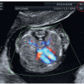
Figure 11.3: Color Doppler of an apical four-chamber view at 12 weeks of gestation by transabdominal (A) and transvaginal (B) ultrasound examination demonstrating diastolic flow from both right (RA) and left (LA) atrium into right (RV) and left (LV) ventricle, respectively. Note that the heart in B is displayed with a higher resolution due to the transvaginal approach.


Figure 11.4: Transvaginal ultrasound of the outflow tracts in color Doppler in a fetus at 13 weeks of gestation showing the five-chamber view (A), the short axis view of the right ventricle (RV) (B), and the three-vessel-trachea view (C). Ao, aorta; LV, left ventricle; PA, pulmonary artery; SVC, superior vena cava.


Figure 11.5: Four essential planes in the first trimester cardiac examination include the plane at the abdominal circumference level (A) to visualize abdominal situs with the stomach (asterisk) on the left side, the four-chamber view (B) in gray scale, as well as the four-chamber view in color Doppler in diastole (C) and the three-vessel-trachea view in color Doppler in systole (D). LV, left ventricle; RV, right ventricle; PA, pulmonary artery; Ao, aorta; R, right; L, Left.


CONGENITAL HEART DISEASE IN THE FIRST TRIMESTER

Congenital heart disease (CHD) is the most common severe congenital abnormality.5,6 About half of the cases of CHD are severe and account for over half of deaths from congenital abnormalities in childhood.5 Moreover, CHD results in the most costly hospital admissions for birth defects in the United States.7 The incidence of CHD is dependent on the age at which the population is initially examined and the definition of CHD used. An incidence of 8 to 9 per 1,000 live births has been reported in large population studies.5,6 It is generally accepted that in the first trimester of pregnancy the prevalence of CHD is higher, as many fetuses with complex anomalies die in utero, especially when associated with extracardiac malformations or early hydrops. One of the poor prognostic signs of CHD is its association with extracardiac anomalies including genetic diseases. The detection of a fetal anomaly is therefore an indication for fetal echocardiography. Even when isolated, CHD can be associated with aneuploidy or syndromic conditions. Prenatal diagnosis of CHD in the first trimester allows for pregnancy counseling and provides enough time for diagnostic options and decision-making.1 CHD can be suspected in the first trimester by the presence of indirect signs such as a thickened nuchal translucency (NT), by the presence of extracardiac malformations, or by the direct observation of cardiac and great vessel anatomic abnormalities.


ASSOCIATED EXTRACARDIAC MALFORMATIONS

Cardiac anomalies are often associated with extracardiac malformations either as part of a defined genetic disease or in isolation. Cardiac anomalies may be found in association with brain, abdominal, urogenital, or skeletal anomalies among others. Even if CHD appears isolated, a careful follow-up should be performed in the second trimester to look for associated extracardiac anomalies. Either isolated or in combination with other extracardiac anomalies, the detection of a CHD is a major hint for a possible association with aneuploidy or other syndromic conditions. The true incidence of CHD association with aneuploidy is unknown but more than 20% of all CHD detected in the first trimester are associated with chromosomal numeric aberrations. This may represent an overestimation given that a significant percentage of CHD in the first trimester is detected after a thickened NT is diagnosed. Aneuploidies associated with CHD in the first trimester include trisomies 21, 18, and 13, as well as with Turner syndrome and triploidy (see Chapter 6 for more details). Other chromosomal anomalies are possible but rather accidental. One major chromosomal anomaly is the association with deletion 22q11, testing for which has to be offered when invasive procedure is performed, especially when a conotruncal anomaly is detected (see below). Deletions and duplications are more commonly detected nowadays given the widespread use of microarray as a diagnostic test following chorionic villous sampling when CHD is diagnosed in the first trimester. Monogenic diseases associated with cardiac defects are usually not detected in early gestation. For more details on genetics of cardiac anomalies, we recommend monographs and review articles on this subject.1,8


Deletion 22q11.2 Syndrome (DiGeorge Syndrome)

Deletion 22q11.2 syndrome (also called DiGeorge syndrome or CATCH 22) is the most common deletion in humans and is the second most common chromosomal anomaly in infants with CHD (second to trisomy 21). It has an estimated prevalence of 1:2,000 to 1:4,000 live births.9 The acronym CATCH-22 was used to describe features of DiGeorge syndrome to include Cardiac anomalies, Abnormal facies, Thymus hypoplasia, Cleft palate, Hypocalcemia, and the microdeletion on chromosome 22.8 Phenotypic abnormalities include cardiac anomalies, mainly outflow tract abnormalities in combination with thymus hypoplasia or aplasia, cleft palate, velopharyngeal insufficiency, and dysmorphic facial features.9 Disorders of the skeleton can affect the limbs and the spine. Mental disorders are found in 30% of the adults with this deletion.8 Diagnosis of this deletion can be achieved by fluorescence in situ hybridization (FISH) technique or with microarray analysis. In an affected fetus or infant, the parental examination reveals, in approximately 6%, an affected parent with subtle signs of this syndrome with a 50% transmission to future offspring.8 Cardiac anomalies that are found in deletion 22q11.2 syndrome primarily include conotruncal anomalies such as an interrupted aortic arch, common arterial trunk (CAT), absent pulmonary valve syndrome, pulmonary atresia with VSD, tetralogy of Fallot (TOF), and conoventricular septal defects.8,10,11 The presence of a right aortic arch either in isolation or in combination with a cardiac anomaly increases the risk for deletion 22q11.2.12 Reports on the detection of deletion 22q11.2 in the first trimester are scarce, but in our opinion, this is primarily due to missed diagnosis of cardiac and extracardiac abnormalities rather than due to the inability to make the diagnosis in the first trimester. Several ultrasound features of deletion 22q11.2 that are seen in the second trimester such as a small thymus,10 a dilated cavum septi pellucidi,13 or polyhydramnios14 are not seen in the first trimester. Facial dysmorphism, as another feature of deletion 22q11.2, is too subtle to be a reliable sonographic feature, even in the second trimester. In the presence of a first trimester cardiac or extracardiac abnormality in the fetus, genetic counseling for invasive diagnostic testing with chorionic villous sampling or amniocentesis is recommended and with the widespread use of microarray, deletion 22q11.2 will be more commonly detected
in early gestation. Figure 11.6 shows a fetus at 13 weeks of gestation with deletion 22q11.2 detected with targeted FISH performed due to the presence of polydactyly and an interrupted aortic arch seen on the first trimester ultrasound.


Figure 11.6: Fetus at 13 weeks of gestation with deletion 22q11. Note in A the presence of a normal facial profile with normal nuchal translucency (NT). Also note in A the presence of hexadactyly (numbers 1-6), shown in hand. In B, the four-chamber view demonstrates a ventricular septal defect (VSD). C: Obtained at the three-vessel-trachea view and shows an interrupted aortic arch (IAA) (arrows). Chorionic villous sampling with targeted FISH confirmed the suspected deletion 22q11. AAO, ascending aorta; DA, ductus arteriosus; LV, left ventricle; PA, pulmonary artery; RV, right ventricle.


INDIRECT SIGNS OF CARDIAC ANOMALIES IN THE FIRST TRIMESTER

Several ultrasound markers associated with an increased risk for CHD have been described in the first trimester and are today part of the indications for an early fetal echocardiography as listed in Table 11.3. Four of these common ultrasound markers are discussed in the following section.


Increased Nuchal Translucency Thickness

In addition to chromosomal anomalies, several reports have noted an association between increased NT and major fetal malformations including cardiac defects (Fig. 11.7). Prospective studies in mixed low- and high-risk screening populations showed a sensitivity of about 21% for a NT >99th percentile. Studies on the association of NT with CHD have shown that the prevalence of major cardiac defects increases exponentially with fetal NT thickness, without an obvious predilection to a specific CHD.15,16 The underlying pathophysiologic mechanism relating the presence of a thickened NT to fetal cardiac defect is not fully understood.


Table 11.3 • Suggested Indications for Fetal Cardiac Imaging in the First Trimester


Maternal indications


Increased risk for aneuploidy (including maternal or paternal balanced translocations)


Maternal poorly controlled diabetes mellitus


Maternal cardiac teratogen exposure Previous child with complex cardiac malformation


Fetal indications


Thickened nuchal translucency


Abnormal cardiac axis


Reverse flow in A-wave of ductus venosus


Tricuspid regurgitation


Extracardiac fetal malformations


Fetal hydrops in the first trimester


Reversed A-Wave in Ductus Venosus

Under normal conditions, ductus venosus (DV) waveforms show a biphasic pattern throughout the cardiac cycle. Abnormal DV waveform pattern is typically characterized by
an absent or reversed A-wave during the atrial contraction phase of diastole (Fig. 11.8A). This flow pattern in the first trimester has been associated with an increased risk of aneuploidy. In chromosomally normal fetuses, abnormal DV waveforms have also been shown to be associated with structural cardiac anomalies.17 The underlying pathophysiologic mechanism linking the reversed DV A-wave to fetal CHD is unclear, but an increased right atrial preload as a result of an increase in volume, pressure, or both in CHD could be one of the underlying mechanisms. Detecting a reversed A-wave in the DV increases the risk for the presence of CHD in the fetus.16


Figure 11.7: Relationship between increased nuchal translucency (NT) thickness and risk of congenital heart disease (CHD) based on a meta-analysis of 12 studies. Note that the prevalence of CHD increases with increased NT thickness. (Adapted from Clur SA, Ottenkamp J, Bilardo CM. The nuchal translucency and the fetal heart: a literature review. Prenat Diagn. 2009;29:739-748; copyright John Wiley & Sons, Ltd., with permission.)


Tricuspid Regurgitation

TR can occur in the fetus at all gestational ages and can be transient. TR at 11 to 13 weeks of gestation is a common finding in fetuses with trisomies 21, 18, and 13, and in those with major cardiac defects.16 TR is found in about 1% of euploid fetuses, in 55% of fetuses with trisomy 21, in one-third of fetuses with trisomy 18 and trisomy 13, and in one-third of those with complex cardiac defects.18 A standardized approach to the diagnosis of TR is important and includes the following (see also Chapter 1): the image is magnified, an apical four-chamber view of the fetal heart is obtained, pulsed-wave Doppler sample volume of 2.0 to 3.0 mm is positioned across the tricuspid valve, and the angle to the direction of flow is less than 30 degrees from the direction of the interventricular septum. A TR is thus diagnosed when it is seen for at least half of systole with a velocity of over 60 cm per second. The detection of TR (Fig. 11.8B) increases the risk for the presence of a complex cardiac defect.


Figure 11.8: A: Pulsed Doppler of ductus venosus (DV) in a fetus at 13 weeks of gestation with a cardiac defect, showing reversed flow in the A-waves (open circle) during atrial contractions. The presence of this pattern suggests an increased risk for associated cardiac abnormalities. B: Pulsed Doppler of the tricuspid valve (long arrow) in a fetus with a tetralogy of Fallot. Note the presence of tricuspid regurgitation on pulsed Doppler (opposing arrows). The presence of tricuspid regurgitation increases the risk for the presence of cardiac abnormalities. S, peak systolic velocity; D, peak diastolic velocity.


Cardiac Axis in Early Gestation

Several studies have established an association between an abnormal cardiac axis and CHD in mid-second and third trimesters of pregnancy and also recently in early gestation. Cardiac axis measurement in the first trimester can be challenging and requires the use of high-definition color in order to clearly delineate the ventricular septum (Fig. 11.9). In a case-control study design, we have recently reported on the fetal cardiac axis in 197 fetuses with confirmed CHD between 11 0/7 and 14 6/7 weeks of gestation, matched with a control group.19 In the control group, the mean cardiac axis was 44.5 ± 7.4 degrees and did not significantly change in early pregnancy.19 In the CHD group, 25.9% of fetuses had cardiac axis measurements within normal limits.19 In 74.1%, the cardiac axis was abnormal. The performance of cardiac axis measurement in detection of major CHD was significantly better than enlarged NT, TR, or reversed A-wave in DV used alone or in combination.19


COMMON FETAL CARDIAC ANOMALIES

In the following sections, we will present CHD that can be diagnosed in the first trimester of pregnancy. For each fetal cardiac abnormality, we will define the abnormality, describe sonographic findings along with optimal planes for diagnosis in the first trimester, and briefly list associated cardiac and extracardiac malformations. Table 11.4 lists abnormal ultrasound findings and corresponding cardiac anomalies in the first trimester of pregnancy. For more detailed information on prenatal cardiac imaging and CHD in the first, second, and third trimesters of pregnancy, we refer the readers to our book on this subject.1


Figure 11.9: Cardiac axis (blue arrows) measurement in two fetuses at 13 weeks of gestation in color Doppler. In fetus A with a normal heart anatomy, the cardiac axis is normal. In fetus B with an atrioventricular septal defect (AVSD) and ventricle disproportion with aortic coarctation (CoA), the cardiac axis is deviated with a wide angle. Cardiac axis is measured in a four-chamber view of the heart by the angle of two lines; the first line starts at the spine (S) posteriorly and ends in mid-chest anteriorly, bisecting the chest into two equal halves, the second line runs through the ventricular septum. RV, right ventricle; LV, left ventricle; L, left.


Hypoplastic Left Heart Syndrome


Ultrasound Findings

In HLHS, the four-chamber view appears abnormal in gray scale and in color Doppler. Cases with a combined mitral and aortic atresia show an absent left ventricle, and can be detected at 12 to 13 weeks of gestation (Figs. 11.11A and 11.12A). In gray scale, the left ventricle appears small or absent in the 4CV (Figs. 11.11A and 11.12A), and color Doppler shows an absence of flow into the left ventricle (Figs. 11.11B and 11.12B). The classic appearance of a single ventricle on gray scale and color Doppler in the first trimester is thus suggestive of HLHS. When suspected on the 4CV in the first trimester, HLHS should be confirmed in the 3VT view, which reveals an enlarged pulmonary artery with a small aortic arch with
reverse flow on color Doppler (Figs. 11.11C and 11.12C). An echogenic globular left ventricle can occasionally be seen in the first trimester in HLHS (Fig. 11.13) and represents left ventricular changes (fibroelastosis), similar to that noted in the second and third trimesters of pregnancy. Of note is that the presence of a “normal” four-chamber view in the first trimester cannot rule out HLHS, as it has been shown to develop between the first and second trimesters of gestation.


Table 11.4 • Abnormal Ultrasound Findings and Suspected Cardiac Anomalies in the First Trimester


Four-chamber view in gray scale and color Doppler


Abnormal cardiac axis (left-sided in TOF, CAT—mesocardia in TGA, DORV, dextrocardia in heterotaxy)


Severe tricuspid insufficiency in Ebstein anomaly


Single ventricle in AVSD, univentricular heart, HLHS, tricuspid atresia with VSD


Ventricle disproportion in coarctation of the aorta, HLHS, HRHS, pulmonary atresia with VSD, mitral atresia and tricuspid atresia


Three-vessel-trachea view in color Doppler


Discrepant great vessel size with forward flow in the small vessel in TOF, coarctation of the aorta, Tricuspid atresia with VSD


Discrepant great vessel size with reversed flow in the small vessel in HLHS, HRHS, PA with VSD


Single large great vessel in CAT, PA with VSD


Single great vessel of normal size in TGA or DORV


Interrupted aortic isthmus in interrupted aortic arch


Aortic arch right-sided to the trachea in right-sided aortic arch with left ductus arteriosus, right-sided aortic arch with right ductus arteriosus, and double aortic arch


TOF, tetralogy of Fallot; CAT, common arterial trunk; TGA, transposition of the great arteries; DORV, double outlet right ventricle; AVSD, atrioventricular septal defect; HLHS, hypoplastic left heart syndrome; VSD, ventricular septal defect; HRHS, hypoplastic right heart syndrome; PA, pulmonary atresia.


Figure 11.10: Schematic drawings of hypoplastic left heart syndrome (HLHS). Note in A the typical features of hypoplastic hypokinetic left ventricle (LV), dysplastic mitral valve, atretic aortic valve, and hypoplastic aorta (Ao). B: The infrequent type of HLHS in the first trimester with dilated, hyperechogenic left ventricle (fibroelastosis), narrowing at the aortic valve level and obstruction to left ventricular outflow, in association with critical aortic stenosis. RA, right atrium; RV, right ventricle; PA, pulmonary artery; LA, left atrium.


Associated Malformations

HLHS is associated with a 4% to 5% incidence of chromosomal abnormalities,20 such as Turner syndrome, trisomies 13 and 18, and others, and when HLHS is suspected in the first trimester, counseling with regard to genetic testing should be performed. Extracardiac malformations have been reported in 10% to 25% of infants with HLHS21 with
associated genetic syndromes, such as Turner syndrome, Noonan syndrome, Smith-Lemli-Opitz syndrome, and Holt-Oram syndrome.21 Fetuses with HLHS may develop growth restriction in the late second and third trimesters of pregnancy probably due to a 20% reduction in combined cardiac output.22 When HLHS is diagnosed in the first trimester, follow-up ultrasound examinations are recommended.


Figure 11.11: Hypoplastic left heart syndrome in a fetus at 13 weeks of gestation demonstrated by transabdominal ultrasound. Note in A the absence of a left ventricle (arrow) in the four-chamber view. In B, color Doppler shows diastolic flow between right atrium (RA) and right ventricle (RV) with absent left ventricular flow. In C, three-vessel-trachea view in color Doppler shows antegrade flow in the pulmonary artery (PA) (blue arrow) and retrograde flow into the aortic arch (AoA) (red arrow). LA, left atrium.


Figure 11.12: Hypoplastic left heart syndrome in a fetus at 13 weeks of gestation demonstrated by transvaginal ultrasound (different fetus than in Fig. 11.11). Note in A the absence of a left ventricle (LV) in the four-chamber view. In B, color Doppler shows diastolic flow between right atrium (RA) and right ventricle (RV) with absent left ventricular flow. In C, three-vessel-trachea view in color Doppler shows antegrade flow in the pulmonary artery (PA) and retrograde flow into the small aortic arch (AoA). Note the increased resolution in the ultrasound images as compared to Figure 11.11 obtained transabdominally. Compare with Figure 11.11. LA, left atrium.


Figure 11.13: Four-chamber view in a fetus with hypoplastic left heart syndrome (HLHS) at 13 weeks of gestation with gray-scale (A) and color Doppler (B) imaging. Note the presence in A of a relatively small echogenic left ventricular (LV) cavity. Color Doppler in B shows absence of mitral inflow during diastole. The presence of an echogenic LV is unusually found in HLHS in the first trimester in comparison with the second trimester. RA, right atrium; RV, right ventricle; LA, left atrium.

Only gold members can continue reading. Log In or Register to continue

Stay updated, free articles. Join our Telegram channel

Oct 14, 2019 | Posted by in ULTRASONOGRAPHY | Comments Off on The Fetal Heart and Great Vessels

Full access? Get Clinical Tree

Get Clinical Tree app for offline access