Chapter 5 The lumbar lordosis and the vertebral canal
The lumbar lordosis
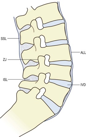
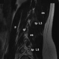
The intact lumbar spine is formed when the five lumbar vertebrae are articulated to one another (Fig. 5.1). Anteriorly the vertebral bodies are separated by the intervertebral discs and are held together by the anterior and posterior longitudinal ligaments. Posteriorly the articular processes form the zygapophysial joints, and consecutive vertebrae are held together by the supraspinous, interspinous and intertransverse ligaments and the ligamenta flava.
Although the lumbar vertebrae can be articulated to form a straight column of vertebrae, this is not the shape assumed by the intact lumbar spine in the upright posture. The reason for this is that the sacrum, on which the lumbar spine rests, is tilted forwards, so that its upper surface is inclined downwards and forwards. From radiographs taken in the supine position, the size of this angle with respect to the horizontal plane of the body has a mean value of about 42–45°,1–3 and is said to increase by about 8° upon standing.1
If a straight lumbar spine articulated with the sacrum, it would consequently be inclined forwards. To restore an upward orientation and to compensate for the inclination of the sacrum, the intact lumbar spine must assume a curve (see Fig. 5.1). This curve is known as the lumbar lordosis.
The junction between the lumbar spine and the sacrum is achieved through joints like those between the lumbar vertebrae. Anteriorly, the body of the L5 vertebra forms an interbody joint with the first sacral vertebra, and the intervertebral disc of this joint is known as the lumbosacral disc. Posteriorly, the inferior articular processes of L5 and the superior articular processes of the sacrum form synovial joints, known either as the L5–S1 zygapophysial joints or as the lumbosacral zygapophysial joints. A ligamentum flavum is present between the laminae of L5 and the sacrum, and an interspinous ligament connects the L5 and S1 spinous processes. However, there is no supraspinous ligament at the L5–S1 level,4 nor are there intertransverse ligaments, the latter having been replaced by the iliolumbar ligament.
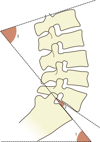
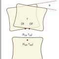
The shape of the lumbar lordosis is achieved as a result of several factors. The first of these is the shape of the lumbosacral intervertebral disc. This disc is unlike any of the other lumbar intervertebral discs in that it is wedge shaped. Its posterior height is about 6–7 mm less than its anterior height.5 Consequently, when the L5 vertebra is articulated to the sacrum, its lower surface does not lie parallel to the upper surface of the sacrum. It is still inclined forwards and downwards but less steeply than the top of the sacrum. The angle formed between the bottom of the L5 vertebra and the top of the sacrum varies from individual to individual over the range 6–29° and has an average size of about 16° (Fig. 5.2).5
The second factor that generates the lumbar lordosis is the shape of the L5 vertebra. Like the lumbosacral disc, the L5 vertebral body is also wedge shaped. The height of its posterior surface is some 3 mm less than the height of its anterior surface.6 As a consequence of the wedge shape of both the L5 body and the lumbosacral disc, the upper surface of L5 lies much closer to a horizontal plane than does the upper surface of the sacrum.
The remainder of the lumbar lordosis is completed simply by inclination of the vertebrae above L5. Each vertebra is inclined slightly backwards in relation to the vertebra below. As a result of this inclination, the anterior parts of the anuli fibrosi and the anterior longitudinal ligament are stretched. Posteriorly, the intervertebral discs are compressed slightly, and the inferior articular processes slide downwards in relation to the superior articular processes of the vertebra below, and may impact either the superior articular process or the pedicle below. The latter phenomenon has particular bearing on the weight-bearing capacity of the zygapophysial joints and is described further in Chapter 8.
Magnitude
Various parameters have been used by different investigators to quantify the curvature of the lumbar lordosis, although they all involve measuring one or other of the angles formed by the lumbar vertebral bodies (see Fig. 5.2). Some have used the angle formed by the planes through the top surface of L1 and the top surface of the sacrum,7,8 and this could be called the ‘L1–S1 lordosis angle’. Fernand and Fox (1985)9 measured the angles between the top of L2 and the top of the sacrum, and between the top of L2 and the bottom of L5, which they called, respectively, the ‘lumbosacral lordotic angle’ and the ‘lumbolumbar lordotic angle’. Others have measured the angle between the top of L3 and the sacrum,10 or the angle formed between planes that bisect the L1–L2 disc and the L5–S1 disc.11,12 Consequently, the measures obtained in these various studies differ somewhat from one another. Nevertheless they all show substantial ranges of variation.
In radiographs taken in the supine position, the angle between the top of L1 and the top of the sacrum varies from 20° to more than 60° but has an average value of about 50°.7 In the standing position, this same angle has been measured as 67° (±3° standard deviation, SD) in children, and 74° (±7° SD) in young males.13 The angle between the top of L2 and the sacrum has a range of 16–80° and a mean value of 45°.9 A value greater than 68° is considered to indicate a hyperlordotic curve.9 However, despite a common belief that excessive lordosis is a risk factor for low back pain, comparison studies reveal that there is no correlation between the shape of the lumbar lordosis and the presence or absence of back pain symptoms.7,10,12
Stability
As described in Chapter 3, the lumbar zygapophysial joints provide a bony locking mechanism that resists forward displacement, and the degree to which a joint affords such resistance is determined by its orientation. The more a superior articular process faces backwards, the greater the resistance it offers to forward displacement.
To resist the tendency for the L5 vertebra to slip forwards, the superior articular processes of the sacrum face considerably backwards. The average orientation of the L5–S1 zygapophysial joints with respect to the sagittal plane is about 45° with most lumbosacral zygapophysial joints assuming this orientation (see Fig. 3.4
Stay updated, free articles. Join our Telegram channel

Full access? Get Clinical Tree


