The imaging findings of some less common diseases of the conal and intraconal compartments* The imaging findings of some less common diseases of the globe* The imaging findings of some less common diseases of the extraconal compartment* • All of the intraocular muscles are usually involved • With advanced disease the lamina papyracea may demonstrate a concavity due to the raised intraorbital pressure • Dynamic contrast-enhanced MRI: the mean of peak enhancement ratio values for the extraocular muscles in Graves’ disease tends to decrease according to the severity of the clinical and anatomical changes
The orbit
THE ORBIT
Within or involving the globe
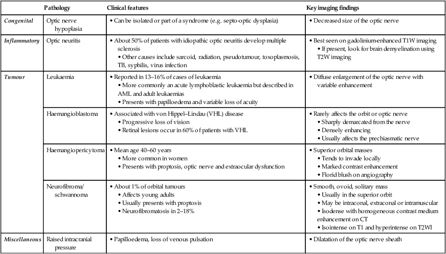
Pathology
Clinical features
Key imaging findings
Congenital
Optic nerve hypoplasia
• Can be isolated or part of a syndrome (e.g. septo-optic dysplasia)
• Decreased size of the optic nerve
Inflammatory
Optic neuritis
• About 50% of patients with idiopathic optic neuritis develop multiple sclerosis
• Other causes include sarcoid, radiation, pseudotumour, toxoplasmosis, TB, syphilis, virus infection
• Best seen on gadolinium-enhanced T1W imaging
• If present, look for brain demyelination using T2W imaging
Tumour
Leukaemia
• Reported in 13–16% of cases of leukaemia
• More commonly an acute lymphoblastic leukaemia but described in AML and adult leukaemias
• Presents with papilloedema and variable loss of acuity
• Diffuse enlargement of the optic nerve with variable enhancement
Haemangioblastoma
• Associated with von Hippel–Lindau (VHL) disease
• Progressive loss of vision
• Retinal lesions occur in 60% of patients with VHL
• Rarely affects the orbit or optic nerve
• Sharply demarcated from the nerve
• Densely enhancing
• Usually affects the prechiasmatic nerve
Haemangiopericytoma
• Mean age 40–60 years
• More common in women
• Presents with proptosis, optic nerve and extraocular dysfunction
• Superior orbital masses
• Tends to invade locally
• Marked contrast enhancement
• Florid blush on angiography
Neurofibroma/ schwannoma
• About 1% of orbital tumours
• Affects young adults
• Usually presents with proptosis
• Neurofibromatosis in 2–18%
• Smooth, ovoid, solitary mass
• Usually in the superior orbit
• May be intraconal, extraconal or intramuscular
• Isodense with homogeneous contrast medium enhancement on CT
• Isointense on T1 and hyperintense on T2WI
Miscellaneous
Raised intracranial pressure
• Papilloedema, loss of venous pulsation
• Dilatation of the optic nerve sheath
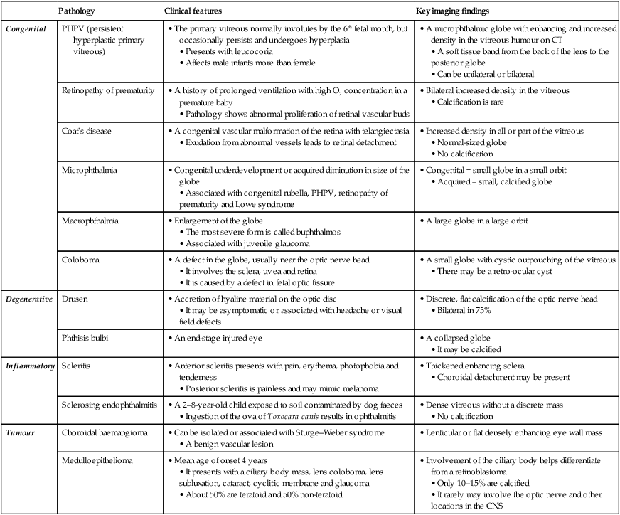
Pathology
Clinical features
Key imaging findings
Congenital
PHPV (persistent hyperplastic primary vitreous)
• The primary vitreous normally involutes by the 6th fetal month, but occasionally persists and undergoes hyperplasia
• Presents with leucocoria
• Affects male infants more than female
• A microphthalmic globe with enhancing and increased density in the vitreous humour on CT
• A soft tissue band from the back of the lens to the posterior globe
• Can be unilateral or bilateral
Retinopathy of prematurity
• A history of prolonged ventilation with high O2 concentration in a premature baby
• Pathology shows abnormal proliferation of retinal vascular buds
• Bilateral increased density in the vitreous
• Calcification is rare
Coat’s disease
• A congenital vascular malformation of the retina with telangiectasia
• Exudation from abnormal vessels leads to retinal detachment
• Increased density in all or part of the vitreous
• Normal-sized globe
• No calcification
Microphthalmia
• Congenital underdevelopment or acquired diminution in size of the globe
• Associated with congenital rubella, PHPV, retinopathy of prematurity and Lowe syndrome
• Congenital = small globe in a small orbit
• Acquired = small, calcified globe
Macrophthalmia
• Enlargement of the globe
• The most severe form is called buphthalmos
• Associated with juvenile glaucoma
• A large globe in a large orbit
Coloboma
• A defect in the globe, usually near the optic nerve head
• It involves the sclera, uvea and retina
• It is caused by a defect in fetal optic fissure
• A small globe with cystic outpouching of the vitreous
• There may be a retro-ocular cyst
Degenerative
Drusen
• Accretion of hyaline material on the optic disc
• It may be asymptomatic or associated with headache or visual field defects
• Discrete, flat calcification of the optic nerve head
• Bilateral in 75%
Phthisis bulbi
• An end-stage injured eye
• A collapsed globe
• It may be calcified
Inflammatory
Scleritis
• Anterior scleritis presents with pain, erythema, photophobia and tenderness
• Posterior scleritis is painless and may mimic melanoma
• Thickened enhancing sclera
• Choroidal detachment may be present
Sclerosing endophthalmitis
• A 2–8-year-old child exposed to soil contaminated by dog faeces
• Ingestion of the ova of Toxocara canis results in ophthalmitis
• Dense vitreous without a discrete mass
• No calcification
Tumour
Choroidal haemangioma
• Can be isolated or associated with Sturge–Weber syndrome
• A benign vascular lesion
• Lenticular or flat densely enhancing eye wall mass
Medulloepithelioma
• Mean age of onset 4 years
• It presents with a ciliary body mass, lens coloboma, lens subluxation, cataract, cyclitic membrane and glaucoma
• About 50% are teratoid and 50% non-teratoid
• Involvement of the ciliary body helps differentiate from a retinoblastoma
• Only 10–15% are calcified
• It rarely may involve the optic nerve and other locations in the CNS
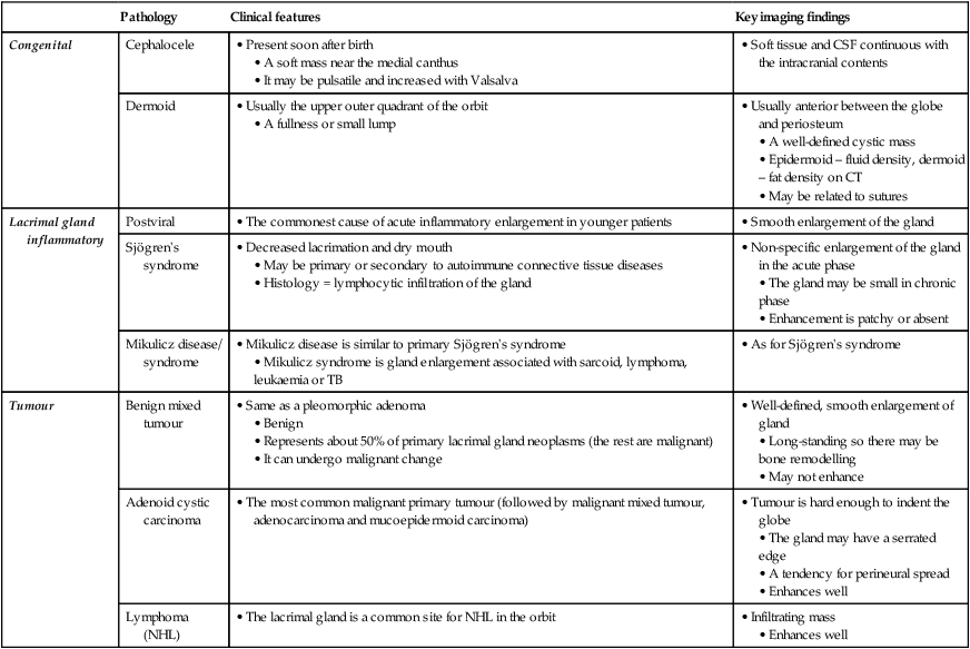
Pathology
Clinical features
Key imaging findings
Congenital
Cephalocele
• Present soon after birth
• A soft mass near the medial canthus
• It may be pulsatile and increased with Valsalva
• Soft tissue and CSF continuous with the intracranial contents
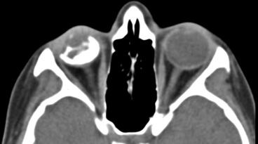
Dermoid
• Usually the upper outer quadrant of the orbit
• A fullness or small lump
• Usually anterior between the globe and periosteum
• A well-defined cystic mass
• Epidermoid – fluid density, dermoid – fat density on CT
• May be related to sutures
Lacrimal gland inflammatory
Postviral
• The commonest cause of acute inflammatory enlargement in younger patients
• Smooth enlargement of the gland
Sjögren’s syndrome
• Decreased lacrimation and dry mouth
• May be primary or secondary to autoimmune connective tissue diseases
• Histology = lymphocytic infiltration of the gland
• Non-specific enlargement of the gland in the acute phase
• The gland may be small in chronic phase
• Enhancement is patchy or absent
Mikulicz disease/ syndrome
• Mikulicz disease is similar to primary Sjögren’s syndrome
• Mikulicz syndrome is gland enlargement associated with sarcoid, lymphoma, leukaemia or TB
• As for Sjögren’s syndrome
Tumour
Benign mixed tumour
• Same as a pleomorphic adenoma
• Benign
• Represents about 50% of primary lacrimal gland neoplasms (the rest are malignant)
• It can undergo malignant change
• Well-defined, smooth enlargement of gland
• Long-standing so there may be bone remodelling
• May not enhance
Adenoid cystic carcinoma
• The most common malignant primary tumour (followed by malignant mixed tumour, adenocarcinoma and mucoepidermoid carcinoma)
• Tumour is hard enough to indent the globe
• The gland may have a serrated edge
• A tendency for perineural spread
• Enhances well
Lymphoma (NHL)
• The lacrimal gland is a common site for NHL in the orbit
• Infiltrating mass
• Enhances well
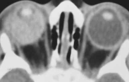
CONAL AND EXTRACONAL DISORDERS
CONAL COMPARTMENT – THYROID OPHTHALMOPATHY
Radiological features
 if there is isolated enlargement of the lateral rectus muscle belly, then causes other than a thyroid ophthalmopathy should be sought (e.g. a pseudotumour)
 The order of muscular involvement: inferior rectus
  medial rectus
  superior rectus
  lateral rectus
  the oblique muscles (‘I’M SLOW’)
 the mean rate of enhancement also decreases according to the disease severity

 metastases
 metastases
 epidermoid
  teratoma
 lymphohaemangioma
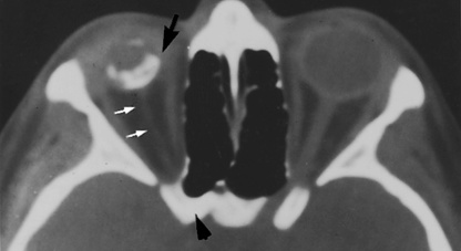
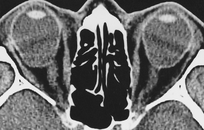
 only 10% of patients are euthyroid
 fusiform enlargement and enhancement of the extraocular muscle bellies (with sparing of the tendinous insertions)
  the hypertrophied muscles and increased fat content may lead to crowding of the orbital apex (with possible optic nerve compression and decreased vision)
 it demonstrates uniform enhancement and is associated with bone destruction