The rotator interval is an anatomically defined triangular area located between the coracoid process, the superior aspect of the subscapularis, and the anterior aspect of the supraspinatus. It is widely accepted that the rotator interval structures fulfill a role in biomechanics and pathology of the glenohumeral joint and long head biceps tendon. However, there is ongoing debate regarding the biomechanical details and the indications for treatment. A better understanding of rotator interval anatomy and function will lead to improved treatment of rotator interval abnormalities, and guide the indications for imaging and surgical intervention.
- •
The rotator interval includes the superior glenohumeral ligament (SGHL) and the coracohumeral ligament (CHL), as well as the long head biceps tendon (LHBT) and the biceps tendon pulley.
- •
The rotator interval ligaments, LHBT, and the adjacent supraspinatus (SST) and subscapularis (SSC) tendon fibers are intimately associated in the lateral portion of the rotator interval.
- •
The SGHL and CHL play a role in glenohumeral joint stability while the bicipital pulley confers LHBT stability.
- •
Rotator interval capsuloligamentous lesions are associated with a spectrum of pathological conditions ranging from glenohumeral instability to adhesive capsulitis.
- •
LHBT lesions and instability are a source of anterior shoulder pain.
- •
LHBT instability patterns are associated with specific patterns of injury to the biceps pulley and adjacent rotator cuff tendons.
- •
Clinical and arthroscopic diagnosis of rotator interval and LHBT abnormality may be difficult.
- •
MR arthrography is well suited to depict rotator interval and LHBT lesions.
- •
Close inspection of the LHBT, biceps pulley, and rotator interval ligaments should be performed if tears of the leading edge of the distal SST tendon or the superior aspect of the distal SSC tendon are identified on magnetic resonance (MR) imaging.
The term rotator interval was first applied by Neer in 1970 when describing greater tuberosity fractures. The dictionary definition of an interval as a space may be misleading, as it pertains to the rotator interval, because “space” implies an area devoid of structures or function. It is widely accepted that the rotator interval structures fulfill a role in biomechanics and pathology of the glenohumeral joint and long head biceps tendon (LHBT). However, there is ongoing debate regarding the biomechanical details as well as the indications for treatment. A better understanding of rotator interval anatomy and function will lead to improved treatment of rotator interval abnormalities, and guide the indications for imaging and surgical intervention.
Anatomy
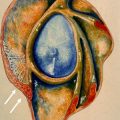
The rotator interval is an anatomically defined triangular area located between the coracoid process, the superior aspect of the subscapularis (SSC), and the anterior aspect of the supraspinatus (SST), with the coracoid process at the base and the bicipital groove at the apex ( Fig. 1 ). The interval is bridged by the glenohumeral capsule, which is composed histologically of loose connective tissue in which there are thick bands of collagen fibers that form the coracohumeral ligament (CHL), superior glenohumeral ligament (SGHL), and the rotator cable. The LHBT traverses the rotator interval as it courses toward the bicipital groove.

The constituents of the rotator interval are closely intertwined and are also associated with the adjacent rotator cuff tendons. While there is a general consensus regarding the contents of the rotator interval as well as the close association between rotator interval ligaments, rotator cuff tendons, and the LHBT, there is a debate regarding their anatomic detail and function. The capsule-ligamentous layer is morphologically the most variable layer of the shoulder, which may explain the variable descriptions of the rotator interval components.
The Ligaments
The CHL and SGHL run a separate course medially but are more intimately associated laterally (see Fig. 1 ). There are contradictory descriptions of the anatomy of the CHL and SGHL, which may relate to differences in study methods emphasizing the intra-articular or extra-articular portion of the ligaments, the use of fresh versus frozen cadavers, and the close association between these ligaments in the mid to lateral portion of the rotator interval.
The CHL is a well-developed structure in most shoulders. This ligament is composed of relatively poorly organized dense connective tissue, and although the existence of the CHL as a true ligament is not consistently supported on histologic studies, it is a constant macroscopic network of fibers or a capsular fold noted in the anterior aspect of the glenohumeral capsule. The CHL arises from the lateral aspect of the coracoid process ; coursing laterally it is inseparable from the capsule, spanning over the rotator interval superficial to the SGHL. Distally this ligament is thought to form 2 major bands, the medial band (MCHL) and the lateral band (LCHL), which are believed to contribute to different components of the ligamentous sling encompassing the LHBT.
The SGHL is a constant but variably expressed anatomic structure composed of parallel bundles of collagen fibers and fibroblasts. Medially, the SGHL has been described as arising from the supraglenoid tuberosity or from the superior labrum at the 1 o’clock position, while Turkel and colleagues also described an additional proximal attachment at the base of the coracoid. The SGHL then courses laterally, parallel to the LHBT, with variable descriptions of the lateral attachment: to the lesser tuberosity, along the bicipital groove floor and over the groove, and merging with the CHL and the humeral semicircular ligament/rotator cable. In a recent cadaveric study, Kask and colleagues described oblique and direct components of the SGHL. The oblique fibers typically arise from the supraglenoid tuberosity and rarely from the anterior superior labrum, fuse loosely with the overlying fibers of the CHL, course over the intra-articular LHBT, and insert on the humeral semicircular ligament/rotator cable. The direct fibers originate from the anterior superior and anterior labrum, run parallel to the LHBT, partially insert on the lesser tuberosity, then course along the floor of the bicipital groove and partially bridge over it. This description of 2 bands with a medial and lateral distal attachment is reminiscent of the 2 distal bands of the CHL. Both superior glenohumeral and CHLs are firmly attached to the outer nonarticular surface of the joint capsule.
The Long Head Biceps Tendon
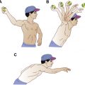
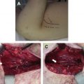
The proximal LHBT is an intra-articular but extrasynovial structure arising from the supraglenoid tuberosity and from the posterior or posterior and superior labrum. The LHBT is flatter and larger in the intra-articular segment with a variable morphology, especially at the origin. Accessory heads of the biceps brachii muscle have been described in approximately 9% to 20% of individuals from the greater tuberosity near the articular capsule, from the articular surface of the glenohumeral joint, from the coracoid process, or in the form of a bifurcated tendon. Variations of the LHBT origin have also been described: LHBT confluent with the rotator cuff, originating from the anterior border of the SST tendon, and as of extra-articular origin. Dierickx and colleagues had described 12 variations of the intra-articular LHBT based on the anatomy and the dynamic behavior during arthroscopy, including variable attachment of the LHBT to the rotator cuff or capsule from a partial mesotenon ( Figs. 2 and 3 ) to a complete adherence of the LHBT to the inferior surface of the capsule, as well as a split biceps tendon and complete absence of the biceps tendon. The authors are of the opinion that these variations are a result of partial detachment from the mesothelium, or synovial fusion with the inferior surface of the capsule, and that some of these variants may cause abnormalities through inferior traction on the SST tendon during abduction.

Related posts:
 Magnetic Resonance Imaging of Rotator Cuff Disease and External Impingement
 Internal Impingement Syndromes
 The Throwing Shoulder: the Orthopedist Perspective
Magnetic Resonance Imaging of the Pediatric Shoulder
The Throwing Shoulder: the Orthopedist Perspective
Magnetic Resonance Imaging of the Pediatric Shoulder
 Entrapment Neuropathies of the Shoulder
 Anatomic Variants and Pitfalls of the Labrum, Glenoid Cartilage, and Glenohumeral Ligaments
Stay updated, free articles. Join our Telegram channel

Full access? Get Clinical Tree


