CHAPTER 94 Thoracic Aortic Trauma
Traumatic injury of the thoracic aorta is a serious and potentially fatal condition. Accurate diagnosis by imaging is essential for expeditious and potentially lifesaving management. CT angiography is the standard imaging technique because of easy availability and a high degree of accuracy. The injury involves a tear of one or more layers of the aortic wall and is usually located at the distal aortic arch or proximal descending thoracic aorta. Key imaging findings include intimal flap, pseudoaneurysm, aortic contour abnormality, and mediastinal hematoma. Intervention is almost always required and consists of either endovascular stent graft or open surgical repair.
THORACIC AORTIC TRAUMA
Etiology and Pathophysiology
Blunt aortic trauma is caused by a severe deceleration injury. The aortic arch is relatively fixed to the thoracic inlet by the brachiocephalic vessels, whereas the ascending and descending aorta are relatively mobile. During deceleration, horizontal shear forces act unequally on the relatively fixed arch and mobile ascending and descending aorta, causing extreme stress on the points of attachment of the aorta, namely, the aortic root and aortic isthmus, rendering these sites susceptible to transection (Fig. 94-1).1 In addition, rapid deceleration may cause the vertebral column, sternum, first rib, and clavicle to compress the aorta in the region of the isthmus, contributing to the mechanism of injury.2
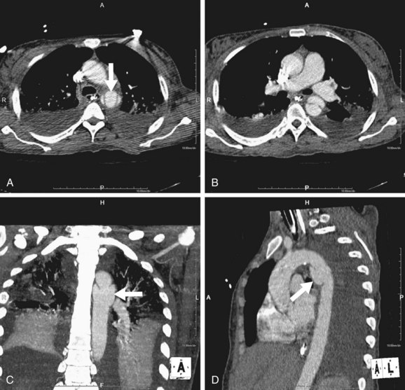
FIGURE 94-1 CT demonstrates traumatic aortic injury at the classic location of the aortic isthmus (arrow) and evidenced by the curvilinear intimal flap (cranial to caudal, A and B). On reformatted images (oblique coronal, C; oblique sagittal, D), the resultant pseudoaneurysm (arrow) is much more easily appreciated.
In clinical practice, the aortic isthmus is by far the most common site of aortic injury; the aortic root and ascending aorta represent 5% or less of cases (Fig. 94-2). However, in autopsy series, the ascending aorta is involved in 25% of cases.3 This discrepancy is explained by the fact that many patients with ascending aortic injury do not survive to reach the hospital or are not stable enough for imaging because of grave complications of ascending aortic trauma, such as hemopericardium with tamponade, disruption of the aortic valve, and coronary artery dissection.
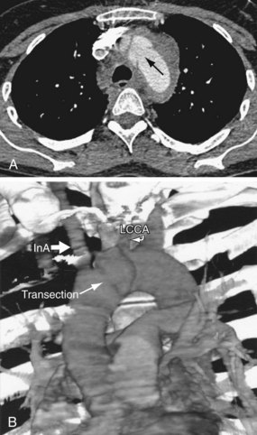
FIGURE 94-2 A, Axial image from multidetector helical CT of the chest in a 40-year-old woman after a high-speed motor vehicle accident demonstrates aortic transection (arrow) with pseudoaneurysm and large periaortic hematoma containing the free aortic rupture. B, Volume rendered reconstruction demonstrates the location of the transection between the takeoff of the innominate artery (InA) and left common carotid arteries (LCCA).
(From Leshnower BG, Litt HI, Gleason TG. Anterior approach to traumatic mid aortic arch transection. Ann Thorac Surg 2006; 81:343-345.)
The degree of traumatic aortic injury depends on the nature of disruption of the aortic wall. Potential aortic injuries range from intimal hemorrhage, through intimal laceration and medial laceration, to complete laceration of the aorta.1 Disruption of the intima and media results in a pseudoaneurysm. Complete laceration, with disruption of the adventitia, usually results in immediate death.
Manifestations of Disease
Clinical Presentation
A history of high-speed deceleration or blunt chest trauma should suggest thoracic aortic injury. Chest or upper back pain, dyspnea, dysphagia, absence of femoral pulses, ecchymoses of the chest wall, seat belt injuries, and fractured sternum act to increase suspicion but are frequently absent.
Imaging Indications and Algorithm
Indications for imaging are based on the mechanism of trauma and clinical signs detailed earlier. As part of a trauma series, most patients will undergo initial chest radiography but will progress immediately to CT regardless of the radiographic results, provided the patient is stable enough.
Imaging Techniques and Findings
Radiography
Plain film findings associated with aortic trauma include a widened mediastinal contour, obscuration of the aortic arch or descending aortic margin, displacement of the trachea or of a nasogastric tube (if present) to the right, and left apical cap (Fig. 94-3). Fracture of the first or second ribs may be seen as a result of the causative trauma.4 However, each of these findings has been shown to have poor sensitivity or poor specificity. For example, in the trauma setting, a widened mediastinum is more commonly explained by anteroposterior positioning, lung contusion, hemothorax, and venous mediastinal bleeding, decreasing the specificity of these findings. Furthermore, in the majority of aortic injuries, the adventitia remains intact, resulting in only mild radiographic mediastinal findings, decreasing the sensitivity of plain radiography. In one study of aortic trauma, 44% of patients with subsequently proven aortic injury had a normal chest radiograph.5 Therefore, plain radiography is not sufficiently sensitive or specific for the evaluation of aortic injury, and in the correct clinical setting, the patient should proceed directly to other modes of imaging.
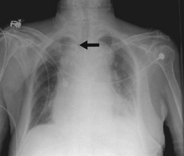
Ultrasonography
Transthoracic ultrasonography has little role in the diagnosis of acute aortic trauma because it would delay CT evaluation, the preferred imaging technique. Transesophageal echocardiography (TEE), on the other hand, has the benefit of portability, which is an advantage for bedside evaluation of very unstable patients. However, in patients with suspected cervical spine injury and facial trauma, TEE is contraindicated. In addition, TEE is highly operator dependent and there is difficulty visualizing the distal ascending aorta and the aortic branch vessels. Therefore, TEE is rarely used clinically for evaluation of aortic trauma, although it may be used in some unusual cases when imaging options are limited or further confirmation is desired.
Computed Tomography
Multidetector computed tomography (MDCT) is the definitive modality for diagnosis of aortic trauma because of its speed, accuracy, postprocessing advantages, and ability to simultaneously evaluate the whole body for traumatic injury. CT angiography has been shown to have 100% sensitivity and 100% negative predictive value for traumatic aortic injury, at lower cost and with fewer complications than by catheter angiography.6
MDCT Technique
In most trauma centers, chest CT is performed as part of a trauma protocol that consists of an unenhanced head CT scan followed by an enhanced study of the chest, abdomen, and pelvis, with use of intravenous contrast material only. Evaluation of the neck can be included if cervical trauma is suspected. The chest CT scan is usually performed in the arterial phase, triggered by bolus timing from the mid-descending aorta. Images are obtained with 1-mm or thinner collimations; 3- to 5-mm axial sections are used for initial review. However, the data should be viewed in multiple planes, which can be achieved either by three-plane reconstructions generated by the CT technologist or by use of an independent workstation by the interpreting physician.
Aortic Pseudoaneurysm
Most patients with aortic trauma who survive to imaging will have a partial tear of the aortic wall. If the intima and media are torn but the adventitia remains intact, blood is forced between the layers, causing formation of a pseudoaneurysm. The imaging appearance is of a rounded bulge projecting from the aorta (Fig. 94-4). The margins are typically irregular, and there is often an intimal flap that forms a “collar” at the neck of the pseudoaneurysm. Note that the injury to the intima may extend several centimeters proximal or distal to the visualized pseudoaneurysm. The extent of the injury, especially the proximal extent, is crucial in the planning of repair because branch vessel involvement has a major influence on repair technique.
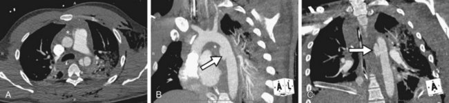
FIGURE 94-4 Axial CT image in a trauma patient demonstrates an intimal flap in the proximal descending thoracic aorta (A). Oblique sagittal maximum intensity projection (B) and oblique coronal (C) reformatted images depict a pseudoaneurysm (arrow). Note extensive mediastinal hematoma (asterisk) diffusely engulfing the aorta, great vessels, and remainder of the mediastinal structures.
Intimal Flap
An intimal flap arises from blood dissecting through an intimal tear as described earlier. It is commonly but not always seen in association with a pseudoaneurysm. To best visualize a flap and to avoid a false-negative diagnosis, thin-section images and an appropriate window must be employed. An intimal flap can be obscured by thick-slab maximum intensity projection images or rendered undetectable by suboptimal windowing during subsequent viewing. Care must be taken to evaluate source or single-voxel multiplanar reconstruction images to ensure that an intimal flap is not present.

Stay updated, free articles. Join our Telegram channel

Full access? Get Clinical Tree

