and Marguerite M. Caré1
(1)
Department of Radiology ML5031, Cincinnati Children’s Hospital, Cincinnati, Ohio, USA
Keywords
Child abusePancreasForeign bodies5.1 Chest/Thorax
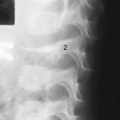
Bony findings include unexpected (or suspected) rib fractures , as well as unexplained fractures of clavicle, acromion and other scapular sites, sternum, and vertebral bodies or spinous processes. Chest radiographs may also reveal traumatic pneumothorax , including responsible foreign bodies ; suspicious foreign bodies in the esophagus; evidence of aspiration forced by a perpetrator; and pneumatocele as consequence of hydrocarbon ingestion aspiration.
Child abuse may lead to involution of the thymus. If the abusive or neglect cause is severe and long-term, immunodeficiency may result [1]. However, remember that congenital immunodeficiency may also have absence of thymus.
Since passive smoking may be considered a form of abuse, the finding of unexplained right middle lobe pneumonia may lead to suspicion of that condition, since such passive smoke exposure may specifically be associated with chronic changes in that lobe.
Flail chest in an infant absent a pertinent trauma history or metabolic bone disease may well be due to abuse.
5.2 Abdominal Organs
Ascites may be due to hemorrhage from solid organ injury; pneumoperitoneum may arise from gastrointestinal tract perforation; retroperitoneal gas or fluid may result from retroperitoneal injury. Rectal perforation may lead to gas or stool or fluid in the pertinent space. With regard to ascites on supine plain images, the centralized bowel loops may be floating anteriorly above ascites, but such centralization may also occur due to greatly enlarged kidneys pushing those loops forward into the central position.
Since hemidiaphragm rupture may be a consequence of abdominal injury, consider whether the trauma might have been abusive.
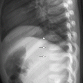
A stomach may be seen dilated with fluid or food if an abused or neglected child is allowed to eat or drink as much as it wishes before coming to radiology for a study (Fig. 5.1). If the child is also small for age, we may be dealing with psychosocial dwarfism. Crying itself often distends the stomach with air.

Fig. 5.1
Stomach greatly distended with food. A 6-year-old boy who was severely neglected and poorly fed for several months. When he arrived at the medical center he was permitted to eat as much as he wished, which he did, before the imaging, which included this abdominal CT that showed massive stomach distention (S)
Occasionally duodenal hematoma may be perceived on plain images by interface with duodenal gas in the duodenum. Ultrasound and other cross-sectional imaging can confirm a suspected traumatic duodenal hematoma. Renal trauma may lead to a perirenal urinoma which can be suspected by an apparent enlarged kidney on plain images.
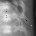
5.2.1 Pancreas
Manifestions of traumatic pancreatitis , especially on cross-sectional imaging, include (Fig. 5.2) discontinuity of the pancreas parenchyma, pancreatic pseudocyst in the organ’s vicinity, peritoneal fluid – sometimes massive, lesser sac location of peritoneal fluid, pleural effusion, and abnormal impression on the inferior border of the transverse colon.