and Marguerite M. Caré1
(1)
Department of Radiology ML5031, Cincinnati Children’s Hospital, Cincinnati, Ohio, USA
Keywords
FractureMetaphysisRibsPeriosteal reaction3.1 Fractures
Even though fractures of the shafts of long bones may well occur from accidental trauma, such fractures in the first year of life, and to some extent in the second year, are often due to abuse, especially in the nonmobile baby. Many statistical reviews, for example [1], have shown a majority of such fractures, notably in femur or humerus, have been found to be abusive. Finding fractures of different ages , such as one without callus (injury less than 10 days old) and another with callus (greater than 10 days old), should increase the suspicion (Figs. 3.1 and 3.2). The orientation of fracture, such as spiral vs. transverse, should not be used as evidence for or against abusive trauma.
Fig. 3.1
Fractures of different ages strongly suggesting abuse. The black lines show the angle from an acute midshaft fracture of the femur in this infant. The lack of periosteal reaction confirms the fracture is less than 9 days old. Meanwhile, the arrow points to periosteal reaction of a healing metaphyseal corner fracture of the tibia (Modified from Oestreich and Crawford [15], with permission)
Fig. 3.2
Fractures of different ages strongly suggesting abuse. In addition to the typical classic metaphyseal lesion corner fracture distally (arrow), this 13-month-old child shows deformity from a healed old midshaft fracture (arrowhead), which was shown acutely on images from 6 months previously (From Oestreich and Crawford [15], with permission)
Fractures of certain bones are particularly suspicious for abuse, including the clavicle (Fig. 3.3) in the first year of life but beyond birth trauma, the acromion (Fig. 3.3), the sternum (look carefully on lateral chest and thoracic spine images), and the first rib (Fig. 3.4). With regard to first rib fractures, do not misdiagnose as a fracture a pseudarthrosis of a cervical rib to a process extending upward from the subjacent rib (Fig. 3.5). This is a hox (homeobox) gene abnormality seen occasionally (perhaps one in 200 persons) [2]. With regard to acromion fractures (Fig. 3.6), the finding of bilateral acromion fractures of the same age may be a consequence of neonatal tetanus [3] and Currarino has described normal acromion lucencies in infancy that simulate fractures [4]. Healing acromion fracture in infancy may show alterations in radiodensity (callus) without frank periosteal reaction.
Fig. 3.3
In this child the healing fractures of the left midclavicle, right acromion, proximal left humerus metaphysis, and proximal right humerus metaphysis strongly suggest abuse. Moreover, periosteal reaction is vigorous at the left humerus and not yet calcified at the right humerus, indicating fractures of different ages
Fig. 3.4
(a) An acute first rib fracture in a baby (arrow) is strongly suspicious for abuse trauma (in absence of metabolic bone disease). (b) Bilateral healing first rib fractures (arrowheads) with periosteal reaction similarly strongly suggest abuse in an infant (in the absence of tetanus)
Fig. 3.5
The fibrocartilaginous gap (arrow) between the left first rib and an upward direct extension of the subjacent rib should not be mistaken for a first rib fracture (Reprinted from Oestreich [2], with permission)
Fig. 3.6
Not only is the left acromion fractured (arrow) in this infant, but also the proximal humerus metaphysis
Fractures of the small tubular bones of the hands and feet in the first year of life should be considered a consequence of abuse (Fig. 3.7), absent metabolic bone disease or, rarely, self-mutulating conditions such as Lesch-Nyhan or Riley-Day syndromes.
Fig. 3.7
This healing buckle fracture (arrowhead) of the proximal first metatarsal was additional evidence of abusive trauma in the subject shown on Fig. 3.3
With questionable fractures, the follow up after more than 10 days may reveal callus/periosteal reaction that confirms the fracture (Fig. 3.8a, b). A delayed finding of deformity, including of a small tubular bone (Fig. 3.9), may signify prior abuse.
Fig. 3.8
(a) The slender sliver of bony density along the medial humeral cortex (arrowhead) was questioned to represent a fracture in this 4-month-old boy. (b) Two and a half weeks later, the vigorous periosteal reaction (arrowhead) leaves no doubt that a fracture had indeed occurred (not immobilized in the interim)
Fig. 3.9
The exostosis-like lateral protrusion of the head of the proximal phalanx of the index finger in this toddler (arrow) raised the question of deformity from a prior abusive fracture. Indeed, the retrieved earlier image from infancy confirmed an acute fracture at that level
Beware of the manubrium centers simulating rib callus on oblique infant chest images, when they overlap (see Fig. 2.13).
3.2 CML (Classic Metaphyseal Lesion s)
One fracture which, in the absence of a compelling other explanation, is strongly suspicious for abuse is widely known as the classic metaphyseal lesion. Corner fractures and bucket handle fractures are both manifestations of the classic metaphyseal lesion, each appearing depending on the angle of the x-ray beam to the fracture (Figs. 3.10 and 3.11). See also Figs. 3.1 and 3.2 for examples of the corner fracture appearance. A CML-like fracture occasionally is a manifestation of childbirth (Fig. 3.12). The use of the follow up at least 10 days later may confirm or rule out a questionable corner fracture (Fig. 3.13).
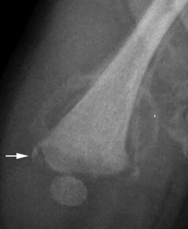
Fig. 3.10
The angle of x-ray beam shows the buckle-handle version of the classic metaphyseal lesion at the proximal tibia (arrow) of this infant who had suffered abusive trauma. The fracture has begun to heal as evidenced by the periosteal reaction (arrowhead) along the tibia shaft, with periosteal reaction as well (arrowhead) along the also fractured fibula
Fig. 3.11
For this healing classic metaphyseal lesion fracture of a proximal humerus, one can see both the metaphyseal corner fracture (arrowhead) and bucket-handle fragment (arrow)
Fig. 3.12
Distal femur metaphyseal corner fragment (arrow) in this 9-day-old with just-beginning-to-calcify periosteal reaction. Birth trauma simulating a classic metaphyseal lesion
Fig. 3.13
(a) A faint corner fragment at the lateral physis of this infant’s proximal humerus (arrow) is a subtle sign of a metaphyseal corner fracture. (b) Follow up a few weeks later confirms the fracture by the vigorous calcified periosteal reaction
3.3 Ten Day Rule: Periosteal Reaction
In general it takes 10 days (well, perhaps 9 days) for periosteal reaction to calcify sufficiently to be visible on conventional radiographs (Fig. 3.14). It takes that long for the matrix vesicles involved in calcification to achieve a proper calcium phosphorus ratio. I think that those investigators who state calcification can occur days earlier err either because of the coincident presence of physiologic periosteal reaction along an involved long bone (which occurs generally between 1 and 6 months of child’s age) or because of improper dating of the traumatic event. A classic description of physiologic periosteal reaction at an equivalent age in puppies was published by Volberg and colleagues [8]. The high incidence of physiologic periosteal reaction in both healthy and sick children was classically commented upon by Shopfner [9].