and Marguerite M. Caré1
(1)
Department of Radiology ML5031, Cincinnati Children’s Hospital, Cincinnati, Ohio, USA
Keywords
Child abuseSkeletal surveyFractures1.1 Observation During Radiography
Before the imaging, be aware of any particular suspected region of injury, either from the purported history or from physical examination by the referring health care practitioners. The technologists (radiographers) or child life colleagues or nursing assistants may be able to observe whether the child interacts normally with the parent or care givers. Indeed, they should observe and relate any suspicions from the behavior of accompanying caregiver. When reviewing skeletal surveys for possible abuse, ask the technologist about any suspicions encountered. Also ask if there was pain or resistance in the course of positioning for a projection.
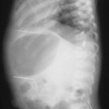
When reviewing the imaging, consider any findings which do not fit with the purported history. Some fractures and other findings are res ipsa loquitur (the thing speaks for itself) suspicious for child abuse, and some become pertinent only when conflicting with that offered history (Fig. 1.1). For example, minor falls, such as from a diaper changing table, are unlikely to cause badly displaced fractures.

Fig. 1.1
The history given by the parents for this infant was “automobile accident.” An acute fracture of the midshaft of the right humerus is shown, as well as a right chest tube and a right pneumothorax. However, fairly mature periosteal reaction is seen along several left ribs (arrowheads), indicating fractures typical for abuse many weeks before. Thus, even if a current accident had occurred, the presence of old healing fractures contradicts the proffered history and thus indicates very likely child abuse
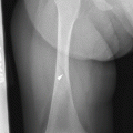
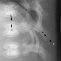
Once the images have been obtained, they should be reviewed by a radiologist while the child is still in the x-ray suite so that additional pertinent views may be obtained; for example, obtain an orthogonal view of a questioned fracture (Fig. 1.2a, b), change the centering for a repeat image of a questioned site (Fig. 1.3), and repeat any inadequate image (Fig. 1.4).


Fig. 1.2
(a) The frontal view of this child’s knee alone barely suggests that anything is amiss. (b) The lateral image reveals a considerably healing fracture from many weeks before. The arrowhead indicates the posterior cortex from before the fracture
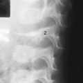
Fig. 1.3
On the original frontal view of the left lower extremity, with x-ray beam centered at the knee, an apparent buckle fracture seems present in the distal tibia diametaphysis. However, a repeat image centered at the ankle shows the “deformity” to actually be the normal metaphyseal collar margin (arrowhead), as defined and discussed in Chap. 2
Fig. 1.4
Motion unsharpness: an example of an ambiguous questionable proximal lateral metaphyseal buckle fracture of the first metatarsal (arrow). Because fractures of small tubular bones in infants are uncommon in the absence of abuse trauma (or metabolic bone disease), it would have been prudent to repeat the image to obtain a view without the motion unsharpness, perhaps with a slightly different obliquity
Related posts:

Stay updated, free articles. Join our Telegram channel
Full access? Get Clinical Tree
