Abstract
FDG avidity in the thymus is often benign thymic rebound. The characteristic triangular shape of benign thymus will distinguish it from malignancy. Thymic malignancies are uncommon, but include thymic carcinomas and malignant carcinoids.
Keywords
FDG, PET/CT, thymus, thymoma, thymic hyperplasia, thymic carcinoma
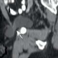
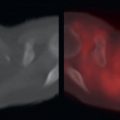
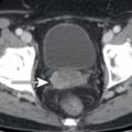
The thymus is a lymphoid organ located anterior to the heart in the anterior mediastinum. Thymic lesions include thymoma, thymic hyperplasia, invasive thymoma, thymic carcinoma, and carcinoid. 18F-fluorodeoxyglucose positron emission tomography/computed tomography (FDG PET/CT) may assist with the characterization of thymic lesions. Thymomas are usually non- to minimally FDG avid ( Fig. 11.1 ), whereas invasive thymomas and thymic carcinomas typically demonstrate substantial FDG avidity ( Fig. 11.2 ). A hallmark feature of invasive thymoma is unilateral pleural metastasis (see Fig. 11.2 ). Following chemotherapy or other systemic therapies, it is common to have rebound thymic hyperplasia. This may be markedly FDG avid and needs to be distinguished from malignancy. The thymus is quite malleable, and the normal thymus is molded into a triangular morphology by the lungs ( Figs. 11.3 and 11.4 ). Contrarily, anterior mediastinal masses are stiffer, form more rounded structures, and subsequently mold the lungs around them. Thus the morphology of an FDG-avid thymus provides substantial value in distinguishing benign from malignant lesions.

Related posts:


Stay updated, free articles. Join our Telegram channel

Full access? Get Clinical Tree


