Abstract
The fetal thymus is a lymphoepithelial structure that grows throughout fetal life and is located in the anterior mediastinum. It is apparent on ultrasound as a homogeneous, quadrangular structure anterior to the major vessels on the three-vessel and three-vessel–trachea view. The thymus can be measured easily in these transverse planes. The finding of thymic hypoplasia or thymic aplasia is clinically important and has been associated with genetic conditions, most commonly 22q11.2 deletion. Thymic hypoplasia is also associated with acquired conditions such as fetal growth restriction and fetal inflammatory response syndrome. A small thymus in fetuses with growth restriction, preterm labor, or preterm premature rupture of membranes may indicate a worse fetal prognosis.
Keywords
fetal thymus, thymic aplasia, thymic hypoplasia, DiGeorge syndrome, 22q11.2 deletion
Introduction
The thymus is a lymphoepithelial organ with key adaptive immune functions during both intrauterine and extrauterine life. The thymus develops from the third pharyngeal pouch, which gives rise to endodermal-derived thymic cortical epithelium, and the third pharyngeal cleft, which is thought to give rise to ectodermal-derived medullary thymic epithelium.
The thymus enlarges in the ninth gestational week when lymphocytes and hematopoietic cells migrate from embryonal blood vessels to spaces between thymus epithelial cells. After the 12th gestational week, the thymus descends into the anterior mediastinum and becomes an encapsulated, lobulated organ with a cortex that is densely populated with lymphocytes and a medulla that appears more epithelial because of a relative paucity of lymphoid cells. The thymus continues to grow throughout fetal life.
Normal Anatomy
General Anatomic Descriptions
The thymus consists of two lateral lobes placed in close contact along the midline, situated partly in the thorax and partly in the neck, and extending from the fourth costal cartilage upward, approaching the lower border of the thyroid gland ( Fig. 6.1 ). It is covered by the sternum and by the origins of the sternohyoid and sternothyroid muscles. Below, it rests on the pericardium, and is separated from the aortic arch and great vessels by a layer of fascia. In the neck, it lies on the anterior surface of the trachea, behind the sternohyoid and sternothyroid muscles.

Ultrasound
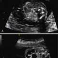
The fetal thymus is visualized on ultrasound (US) as a homogeneous quadrangular structure in the anterior superior mediastinum, ventral to the pericardium and great vessels of the heart, and between both lungs. It is well visualized in the three-vessel view or the three-vessel–trachea view, lying anterior to the vessels (pulmonary artery, aorta, and superior vena cava, with the trachea to the right of the vessels) ( Figs. 6.2–6.5 ). It is most often identified between the sternum and great vessels of the heart in the axial view of the fetal chest. From the sagittal view, the thymus appears triangular or teardrop-shaped ( Fig. 6.6 ). Compared with the lungs, it is similar in echogenicity or slightly more echogenic in the early second trimester, between 17 weeks’ and 22 weeks’ gestation (see Figs. 6.2 and 6.4 ), and less echogenic in later pregnancy (see Figs. 6.3 and 6.5 ).


With the development and application of three-dimensional (3D) US and four-dimensional (4D) US, the entire fetal thymus can be reconstructed and measured using a 3D model derived from multiple two-dimensional (2D) planes, which conquers the limitation of single-plane information from 2D US. Using off-line virtual organ computer-aided analysis (VOCAL) software, the 3D morphology of the fetal thymus can be evaluated from any orientation ( Figs. 6.7–6.9 ).


