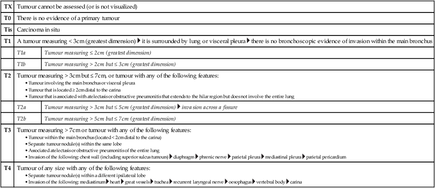
• Tumour within the main bronchus (located < 2cm distal to the carina) • Separate tumour nodule(s) within the same lobe • Associated atelectasis or obstructive pneumonitis of the entire lung • Invasion of the following: chest wall (including superior sulcus tumours)
TNM staging of common cancers
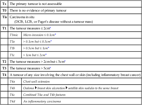
BREAST CANCER
Tx
The primary tumour is not assessable
T0
There is no evidence of primary tumour
Tis
Carcinoma in situ
(DCIS, LCIS, or Paget’s disease without a tumour mass)
T1
The tumour measures ≤ 2cm*
T1mic
Micro-invasion ≤ 0.1cm*
T1a
> 0.1cm but ≤ 0.5cm*
T1b
> 0.5cm but ≤ 1cm*
T1c
> 1cm but ≤ 2cm*
T2
The tumour measures > 2cm but ≤ 5cm*
T3
The tumour measures > 5cm*
T4
A tumour of any size involving the chest wall or skin (including inflammatory breast cancer)
T4a
Chest wall extension
T4b
Oedema  breast skin ulceration
  satellite skin nodules to the same breast
T4c
Combined T4a and T4b features
T4d
An inflammatory carcinoma
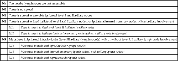
Nx
The nearby lymph nodes are not assessable
N0
There is no spread
N1
There is spread to movable ipsilateral level I and II axillary nodes
N2
There is spread to fixed ipsilateral level I and II axillary nodes, or ipsilateral internal mammary nodes without axillary involvement
N2a
There is spread to fixed level I and II ipsilateral axillary nodes
N2b
There is spread to ipsilateral internal mammary nodes without axillary node involvement
N3
Metastases in ipsilateral infraclavicular (level III axillary) lymph node(s) with or without level I, II axillary lymph node involvement
N3a
Metastases in ipsilateral infraclavicular lymph node(s).
N3b
Metastases in ipsilateral internal mammary lymph node(s) and axillary lymph node(s)
N3c
Metastases in ipsilateral supraclavicular lymph node(s)
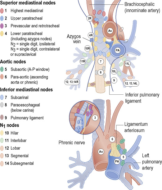
LUNG CANCER
TX
Tumour cannot be assessed (or is not visualized)
T0
There is no evidence of a primary tumour
Tis
Carcinoma in situ
T1
A tumour measuring < 3cm (greatest dimension)  it is surrounded by lung or visceral pleura
  there is no bronchoscopic evidence of invasion within the main bronchus
T1a
Tumour measuring ≤ 2cm (greatest dimension)
T1b
Tumour measuring > 2cm but ≤ 3cm (greatest dimension)
T2
Tumour measuring > 3cm but ≤ 7cm, or tumour with any of the following features:
T2a
Tumour measuring > 3cm but ≤ 5cm (greatest dimension)  invasion across a fissure
T2b
Tumour measuring > 5cm but ≤ 7cm (greatest dimension)
T3
Tumour measuring > 7cm or tumour with any of the following features:
 diaphragm
  phrenic nerve
  parietal pleura
  mediastinal pleura
  parietal pericardium
T4
Tumour of any size with any of the following features:
Nx
The lymph nodes are not assessable
N0
There is no regional nodal involvement
N1
There are involved ipsilateral peribronchial (± ipsilateral) hilar and intrapulmonary lymph nodes
N2
There are involved ipsilateral mediastinal (± subcarinal) lymph nodes
N3
There are involved contralateral mediastinal or hilar nodes, ipsilateral or contralateral scalene nodes, or supraclavicular nodes
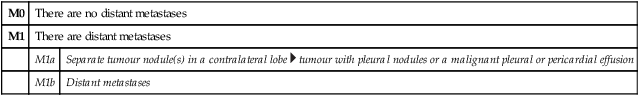
M0
There are no distant metastases
M1
There are distant metastases
M1a
Separate tumour nodule(s) in a contralateral lobe  tumour with pleural nodules or a malignant pleural or pericardial effusion
M1b
Distant metastases
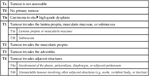
OESOPHAGEAL CANCER
Tx
Tumour is not assessable
T0
No primary tumour
Tis
Carcinoma in situ  high-grade dysplasia
T1
Tumour invades the lamina propria, muscularis mucosae, or submucosa
T1a
Lamina propria or muscularis mucosae
T1b
Submucosa
T2
Tumour invades the muscularis propria
T3
Tumour invades the adventitia
T4
Tumour invades adjacent structures
T4a
Involvement of the pleura, pericardium, diaphragm, or adjacent peritoneum
T4b
Unresectable tumour involving other adjacent structures (e.g. aorta, vertebral body, or trachea)
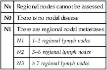
Nx
Regional nodes cannot be assessed
N0
There is no nodal disease
N1
There are regional nodal metastases
N1
1–2 regional lymph nodes
N2
3–6 regional lymph nodes
N3
≥ 7 regional lymph nodes
M0
There are no tumour metastases
M1
There is distant spread (spread of the cancer to organs or distant lymph nodes)
STOMACH CANCER
Tx
Primary tumour is not assessable
T0
There is no evidence of primary tumour
Tis
Carcinoma in situ
T1
Tumour invades the lamina propria or submucosa
T1a
Invasion of the lamina propria or muscularis mucosae
T1b
Invasion of the submucosa
T2
Tumour invades the muscularis propria
T3
Tumour invades the subserosa without invading any adjacent structures
T4
Tumour perforates the serosa or invades adjacent structures
T4a
Perforation of the serosa (visceral peritoneum)
T4b
Invades adjacent structures

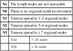
Nx
The lymph nodes are not assessable
N0
There is no regional nodal involvement
N1
Tumour spread to 1–2 regional nodes
N2
Tumour spread to 3–6 regional nodes
N3
Tumour spread to ≥ 7 regional nodes
N3a
7–15 nodes
N3b
≥ 16 nodes
RECTAL CANCER
Tx
Primary tumour is not assessable
T0
There is no evidence of a primary tumour
Tis
Carcinoma in situ: intraepithelial or invasion of lamina propria
T1
Tumour invades the submucosa
T2
Tumour invades, but does not penetrate, the muscularis propria
T3
Tumour invades the subserosa (through the muscularis propria)  there is no involvement of the neighbouring tissues or organs
T3a
The tumour extends < 1mm beyond the muscularis propria
Related posts:
![]()
Stay updated, free articles. Join our Telegram channel

Full access? Get Clinical Tree

 Get Clinical Tree app for offline access

 heart
heart  great vessels
  trachea
  recurrent laryngeal nerve
  oesophagus
  vertebral body
  carina