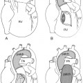
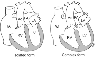
24 Transpositions of the Great Arteries Fig. 24.1 Complete transposition. Asterisk indicates subpulmonary outflow tract, which is often narrowed in the complex form. Ao, ascending aorta; d, ventricular septal defect; LA, left atrium; LV, left ventricle; PA, pulmonary artery; RA, right atrium; RV, right ventricle.
 Transposition is a term to describe ventriculoarterial connection. It refers to discordant ventriculoarterial connection: the right ventricle connects to the aorta and the left ventricle to the pulmonary artery. Transposition occurs with various types of atrioventricular connections:
 There are two classic types of transposition:
 Complete Transposition
Definition and Classification (Fig. 24.1)
 Characterized by normal atria, normally related ventricles, normal atrioventricular connections, and abnormal discordant ventriculoarterial connections
 Also called D-transposition, as it occurs with D-loop ventricles (the morphologically right ventricle on the right side of the morphologically left ventricle) and, in the majority of cases, with a right-sided and anterior aorta. It is a confusing term because of variations in ventricular loop and great arterial relationship.
 Complete transposition as a simple form: about 50% of patients are without associated defects other than a patent foramen ovale or a small patent ductus arteriosus.
 Associated lesions in 50%: ventricular septal defect, left ventricular outflow tract obstruction, and obstructive lesion of the aortic arch.
Pathophysiology
 Concordant atrioventricular and discordant ventriculoarterial connections result in two isolated parallel circulations:
 Survival depends on mixing between these circulations, which may be across a patent foramen ovale or atrial septal defect, additional ventricular septal defect, or patent ductus arteriosus.
 As the pulmonary vascular resistance falls postnatally, there is increased pulmonary blood flow unless an associated pulmonary stenosis is present.
Clinical Manifestations
 Varies according to the presence of associated defects
 Soon after birth with closure of ductus arteriosus there is severe hypoxemia.
 Cyanosis is the most common presentation and is often seen within the first hours of life.
 Mild cyanosis with associated lesions that permit good mixing
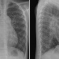
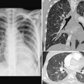
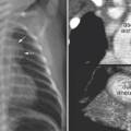
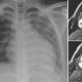
Chest Radiographic Findings
 Neonates often have normal heart size and pulmonary vascularity.
 Increased pulmonary vasculature with an overcirculation pattern with decreasing pulmonary vascular resistance
 Over time, heart size enlarges.
 An oblong egg-on-side appearance due to rightward displacement of the right ventricular outflow tract and combined right atrial and left ventricular dilatation
 Narrow vascular pedicle due to parallel course of anteroposteriorly related great arteries and small thymus as it atrophies with postnatal stress
 Decreased pulmonary vasculature with associated severe pulmonary stenosis
 Aortic arch usually left sided
Pearls
 Most common cyanotic lesion presenting in newborn period
 Classic findings seen over time with decreasing pulmonary vascular resistance
Related posts:
![]()
Stay updated, free articles. Join our Telegram channel

Full access? Get Clinical Tree


Radiology Key
Fastest Radiology Insight Engine