Trauma and Trauma-Related Conditions
COMPLEX REGIONAL PAIN SYNDROME (CRPS)/REFLEX SYMPATHETIC DYSTROPHY SYNDROME (RSDS)
Known also as reflex sympathetic dystrophy syndrome and Sudeck atrophy, CRPS is a clinical syndrome of variable course and unknown cause, characterized by disabling pain, swelling, and vasomotor dysfunction of an extremity. CRPS represents a vastly overdiagnosed illness. There are no specific diagnostic tests, and the diagnosis is based on clinical history, clinical examination, and supportive laboratory findings. There is also no single pathophysiologic mechanism that can explain the diversity and heterogeneity of the symptoms. It can occur following trauma that includes a neurologic injury, and the patient may present with pain and hyperesthesia, diffuse soft tissue swelling, joint stiffness, vasomotor instability, and dystrophic skin changes. There are no acceptable classifications. In 1993, International Association for Study of Pain (IASP) designated a task force to review the nomenclature and develop diagnostic criteria of CRPS. In 1994, at a consensus workshop in Florida, the task force developed the nomenclature (CRPS) and proposed the diagnostic criteria. Furthermore, they subdivided CRPS into types I and II. In CRPS type I, there is no clinically significant neurologic injury, whereas CRPS type II is a classic abnormality first described during civil war that is characterized by well-defined neurologic injury. Some researches have even claimed that the symptoms can spread throughout the body, a claim neither with scientifically proven data nor, even more importantly, with any biologic or medical plausibility. Occasionally, CRPS may be mistakenly diagnosed as arthritis, which can lead to overprescription of pain medication.
Three stages of this condition have been identified. The initial (or acute) inflammatory stage lasts from 1 to 7 weeks and is characterized by diffuse regional pain, inflammation, edema, and hypothermia or hyperthermia. In the second (or dystrophic) stage, which lasts from 3 to 24 months, the clinical findings include pain on exercise, increased sensitivity of the skin to pressure and temperature changes, and skin and muscle atrophy. In the final (or atrophic) stage, irreversible scleroderma-like skin changes and aponeurotic and tendinous retraction may occur.
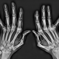
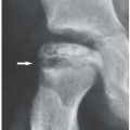
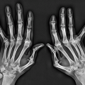
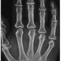
On the radiographs, RSDS is characterized by soft tissue swelling and severe, patchy osteoporosis that progresses rapidly (Fig. 12.1). Bone densitometry shows a lowered bone mineral density content in affected limb. Three-phase technetium bone scan may show increased blood flow, blood pool, and periarticular increased uptake in the affected areas (Fig. 12.2), although these findings are not specific and can be present in patients with other bone abnormalities. The above-listed findings are seen in ˜ 60% of affected patients that truly have CRPS.
The clinical management of patients with CRPS is a nearly daily challenge to rheumatologists, neurologists, neurosurgeons, orthopedic surgeons, and pain specialists. The aim of the treatment is pain control and recovery of limb function. Physical therapy is the first line of treatment. Pharmacologic therapy and selection of the drugs are determined by the severity of the pain and include NSAIDs and COX-2 inhibitors. Metamizol and controlled release opioids (such as hydrocodone or oxycodone) are considered in patients with more severe pain that is not responding to other measures. Recently, neuromodulation of central pain pathways by delivery of an electrical current or chemical application to the central neural axis has been tried with promising results.
TRANSIENT REGIONAL OSTEOPOROSIS
Transient regional osteoporosis is a collective term for a group of conditions that have one feature in common: rapidly developing osteoporosis that usually affects the periarticular regions and has no definite etiology such as trauma or immobilization. It is a self-limited and reversible disorder, of which three subtypes have been described. Transient osteoporosis of the hip is seen predominantly in pregnant women and in young and middle-aged men. Its primary manifestation is local osteoporosis involving the femoral head, femoral neck, and the acetabulum (Fig. 12.3). Regional migratory osteoporosis, which affects the knee, the ankle, and the foot, is mainly seen in men in their fourth and fifth decades. This migratory condition is characterized by pain and swelling around the affected joints and can mimic arthritis. It develops rapidly and subsides in about 6 to 9 months; there may be subsequent recurrence and involvement of other joints. Idiopathic juvenile osteoporosis is commonly seen during or just before puberty and typically regresses spontaneously. Skeletal involvement is often symmetrical and osteoporosis is generally juxta-articular in location. Spine is occasionally affected, and the vertebral body compression fractures may be encountered.
POSTTRAUMATIC MYOSITIS OSSIFICANS
Occasionally, after a fracture, dislocation, or even minor trauma to the soft tissues, an enlarging, well-circumscribed painful mass develops at the site of injury. Clinically, pain and swelling at this site persist for many days. The pain tends to become more localized and may increase in intensity for up to 4 to 6 weeks. The characteristic feature of this lesion includes the clearly recognizable pattern of its evolution, which correlates well with the lapse of time after the trauma. Thus, by the 3rd or 4th week, calcifications and ossifications in the mass begin to develop (Fig. 12.4), and by the 6th to 8th week, the periphery of the mass shows definite, well-organized cortical bone (Fig. 12.5). The important radiographic hallmark of this complication is the presence of the so-called zonal phenomenon. On the radiograph, this phenomenon is characterized by a radiolucent area in the center of the lesion, indicating the formation of an immature bone, and by a dense zone of mature ossification at the periphery (myositis ossificans circumscripta). In addition, a thin radiolucent cleft separates the ossific mass from the adjacent cortex (Fig. 12.6). These important features help differentiate this condition from juxtacortical osteosarcoma, which
may, at times, appear very similar. It must be stressed, however, that occasionally, the focus of myositis ossificans may adhere and fuse with the cortex, mimicking parosteal osteosarcoma on radiographs. In these cases, CT may provide additional information, such as the presence of the zonal phenomenon characteristic of myositis ossificans (Fig. 12.7). The MRI appearance of myositis ossificans depends on the stage of maturation of the lesion. In the early stage, T1-weighted sequences usually show a mass that lacks definable borders with homogeneous intermediate signal intensity, slightly higher than that of adjacent muscle. T2-weighted images show the lesion to be of high signal intensity. The more mature lesions show intermediate signal intensity on T1-weighted sequences isointense with adjacent muscle, surrounded by a rim of low signal intensity, which corresponds to peripheral bone maturation. On T2 weighting, the lesion is generally of high signal intensity but may appear heterogeneous. The rim of low signal is seen at the periphery. Sometimes, the focus of myositis ossificans (whether immature or mature) may contain a fatty component, giving the lesion a high-intensity signal on T1-weighted images (Fig. 12.8). After an intravenous administration of gadopentetate dimeglumine, T1-weighted images show a well-defined peripheral rim of contrast enhancement, but the center of the lesion does not enhance (Fig. 12.9).
may, at times, appear very similar. It must be stressed, however, that occasionally, the focus of myositis ossificans may adhere and fuse with the cortex, mimicking parosteal osteosarcoma on radiographs. In these cases, CT may provide additional information, such as the presence of the zonal phenomenon characteristic of myositis ossificans (Fig. 12.7). The MRI appearance of myositis ossificans depends on the stage of maturation of the lesion. In the early stage, T1-weighted sequences usually show a mass that lacks definable borders with homogeneous intermediate signal intensity, slightly higher than that of adjacent muscle. T2-weighted images show the lesion to be of high signal intensity. The more mature lesions show intermediate signal intensity on T1-weighted sequences isointense with adjacent muscle, surrounded by a rim of low signal intensity, which corresponds to peripheral bone maturation. On T2 weighting, the lesion is generally of high signal intensity but may appear heterogeneous. The rim of low signal is seen at the periphery. Sometimes, the focus of myositis ossificans (whether immature or mature) may contain a fatty component, giving the lesion a high-intensity signal on T1-weighted images (Fig. 12.8). After an intravenous administration of gadopentetate dimeglumine, T1-weighted images show a well-defined peripheral rim of contrast enhancement, but the center of the lesion does not enhance (Fig. 12.9).
The histopathologic features are pathognomonic and consist of the least mature tissue in the center of the lesion and more mature at the periphery, corresponding to the radiographic zonal phenomenon. In the central portion of the lesion, noted are increased cellularity and the presence of immature fibroblastic cells, whereas at the periphery, microtrabeculae with peripheral appositional osteoblasts are present (Fig. 12.10). Biopsy of the lesion in its early stage may lead to erroneous diagnosis of malignancy.
The treatment of myositis ossificans varies with each case. In most cases, so-called “wait and watch” approach is advisable, because the lesion may shrink in time and became asymptomatic. Complete surgical excision is performed after full maturation of the lesion. Nonsurgical treatment with shock waves has been occasionally tried.
POSTTRAUMATIC OSTEOLYSIS OF THE DISTAL CLAVICLE
After injury to the shoulder, such as sprain of the acromioclavicular joint, resorption of the distal (acromial) end of the clavicle may occasionally occur. At times, this abnormality may be mistaken for resorption of the acromial end of the clavicle occurring in rheumatoid arthritis or in the primary hyperparathyroidism. The osteolytic process, which is associated with mild-to-moderate pain, usually begins within 2 months after injury. The initial radiographic findings consist of soft tissue swelling and periarticular osteoporosis associated with slightly irregular contour of acromial end of the clavicle (Fig. 12.11). Later, small erosions may develop (Fig. 12.12). MRI findings comprise increased signal intensity on water-sensitive sequences of the acromial end of the clavicle related
to bone marrow edema, marginal irregularity of the bone outline, and fluid within the acromioclavicular joint (Fig. 12.13). In its late stage, resorption of the distal end of the clavicle results in marked widening of the acromioclavicular joint (Fig. 2.14).
to bone marrow edema, marginal irregularity of the bone outline, and fluid within the acromioclavicular joint (Fig. 12.13). In its late stage, resorption of the distal end of the clavicle results in marked widening of the acromioclavicular joint (Fig. 2.14).
SHOULDER IMPINGEMENT SYNDROME
Impingement syndrome of the shoulder refers to a condition in which the supraspinatus tendon and subacromial bursa are chronically entrapped between the humeral head inferiorly and the anterior acromion itself, spurs of the anterior acromion or acromioclavicular joint, or the coracoacromial ligament (coracoacromial arc) superiorly. Early diagnosis and treatment of impingement syndrome are critical to improve shoulder function and prevent the progression of this condition. Frequently, however, clinical signs and symptoms are nonspecific and may mimic arthritis, and the diagnosis is often delayed until a full-thickness tear in the rotator cuff has developed. Only rarely it can be definitely diagnosed based on the clinical findings, characterized by severe pain during abduction and external rotation of the arm. More reliable are imaging findings associated with this abnormality, including subacromial proliferation of bone, spurring at the inferior aspect of the acromion, and degenerative changes of the humeral tuberosities at the insertion of the rotator cuff.
Neer described three progressive stages of impingement syndrome apparent clinically and at surgery. Stage I consists of edema and hemorrhage and is reversible with conservative therapy. It typically occurs in young individuals engaged in sport activities requiring excessive use of the arm above the head (i.e., swimming). Stage II consists of fibrosis and thickening of the subacromial soft tissues, rotator cuff tendinitis, and sometimes a partial tear of the rotator cuff. It is manifested clinically by recurrent pain and is often seen in patients 25 to 40 years old. Stage III represents complete rupture of the rotator cull and is associated with progressive disability. It is usually seen in patients older than 40 years of age. Because of its high soft tissue contrast resolution and multiplanar imaging capabilities, MRI is the only technique that can accurately image the early changes of this condition, in particular bursal thickening and effusion (subacromial bursitis), edema, and inflammatory changes of the rotator cuff and its tendons (Fig. 12.15A, B), and later changes consisting of partialand full-thickness tear of the rotator cuff (Fig. 12.15C, D).
ULNAR IMPINGEMENT SYNDROME
Stay updated, free articles. Join our Telegram channel

Full access? Get Clinical Tree


