Traumatic Disc Herniation—Adolescent


Clinical Presentation


The patient is a 19-year-old college basketball player with a 4-month history of progressively worsening low back pain, radiating into the thoracic region and also into the right buttock and thigh. The pain is not relieved by rest, aspirin, or nonsteroidal anti-inflammatory agents. Back strengthening exercises over the past 6 weeks have not relieved his pain whatsoever. He reports a tingling sensation in both feet when recumbent at night. No lower extremity weakness on examination.


Imaging Presentation


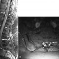
Magnetic resonance (MR) imaging of the lumbar spine reveals evidence of L4-5 intervertebral disc degeneration and a moderate-sized midline L4-5 disc herniation that causes ventral thecal sac deformity ( Figs. 26-1 and 26-2 ) . A lumbar spine computed tomography (CT) scan was obtained because of deformity of the posteroinferior corner of the L4 vertebral body and probable avulsion fracture of posterior margin of the L4 vertebral body seen on the MR scan. The CT scan confirms an avulsion fracture of the posteroinferior corner of the L4 vertebral body associated with L4-5 disc herniation ( Figs. 26-3 and 26-4 ) .


Figure 26-1


Traumatic Disc Herniation L4-5.

19-year-old basketball player with vertebral apophysis avulsion. A herniated disc ( HD ) at L4-5 is demonstrated on sagittal T1-weighted MR image A and sagittal T2-weighted image B . Note deformity of the posteroinferior margin of the L4 vertebral body ( small arrow in image A and B ). Avulsed apophyseal cortical bone fragment ( F ) is T2 hypointense on sagittal T2-weighted image B , but difficult to visualize on sagittal T1-weighted image A . The posterior longitudinal ligament ( P ) is displaced posteriorly by the avulsed bone fragment. Note diminished intradiscal T2 signal intensity in L4-5 intervertebral disc on image B , consistent with disc degeneration.


Figure 26-2


Traumatic Disc Herniation L4-5.

Same patient as in Figure 26-1 . A focal midline herniated disc ( HD ) causes posterior displacement of a T1 and T2 hypointense bone fragment ( F ) as seen on axial T1-weighted MR image A and T2-weighted image B . The herniated disc causes ventral effacement of the thecal sac.


Figure 26-3


Apophyseal Avulsion Fracture.

Same patient as in Figures 26-1 and 26-2 . Sagittal CT image confirms presence of apophyseal avulsion fracture. The herniated disc ( HD ) has caused an avulsion fracture ( F ) of the posterior L4 vertebral cortex by disrupting the posteroinferior vertebral apophysis. An irregular vertebral endplate defect ( D ) is demonstrated where the inferior L4 endplate joins the vertebral apophysis.


Figure 26-4


Apophyseal Avulsion Fracture.

Same patient as in Figures 26-1 to 26-3 . Apophyseal avulsion fracture fragments ( F ) are displaced posteriorly by the herniated disc ( HD ) as demonstrated on contiguous axial CT images A and B obtained just above the L4-5 intervertebral disc level. Image B is slightly superior to image A .


Discussion


In children and adolescents, traumatic disc herniations usually involve the unused ring apophysis. Traumatic disc herniations and ring apophyseal injuries in young persons more commonly occur in the lumbar region, although occasionally they may occur in the cervical or thoracic spine. The injury most commonly involves the inferior rim of the L4 endplate or the superior rim of the S1 endplate ( Figs. 26-1 to 26-8 ) . Most traumatic herniations in children or adolescents occur in athletes or those who perform activities that subject the spine to repetitive stress. Some have suggested that there may be a congenital insufficiency of the ring apophysis that predisposes one to this injury. Motor vehicle accidents or other acute traumatic events can also damage the ring apophysis.


Figure 26-5


Traumatic Disc Herniation L5-S1.

17-year-old patient actively involved in wrestling and weightlifting. Two-month history of severe low back pain and right leg pain. Traumatic disc herniation ( long arrow ) associated with posterosuperior endplate defect ( short arrow ) is demonstrated on sagittal T1-weighted MR image A and T2-weighted image B . The disc herniation ( long arrow ) causes mild ventral thecal sac deformity. Diminished intradiscal T2 signal intensity in L5-S1 disc due to disc degeneration is shown in image B .


Figure 26-6


Traumatic Disc Herniation L5-S1.

Axial T1-weighted MR image A and T2-weighted image B in same patient as in Figure 26-5 . The focal herniated disc ( HD ) is centered to the right of midline, encroaches on the ventral epidural fat, and causes posterior displacement of right S1 nerve root ( short arrow ).


Figure 26-7


Traumatic Disc Herniation L5-S1.

Sagittal post discogram CT in same patient as in Figure 26-6 . Following contrast agent ( C ) injected into the L5-S1 intervertebral disc, the patient reported severe low back pain highly concordant with his usual pain in distribution and intensity. The injected intradiscal contrast agent ( C ) extends into the defect ( D ) in the posterior aspect of the superior S1 endplate. A bone fragment ( F ) is displaced from the vertebral apophysis posteriorly. The herniated disc is outlined posteriorly by anterior epidural contrast ( EDC ) that leaked from the intervertebral disc through a fissure in the posterior annulus.


Figure 26-8


Traumatic Disc Herniation L5-S1.

Same patient as in Figure 26-7 . Axial post discogram CT image A obtained at the disc level and CT image B obtained slightly below the disc level. On image A , the intradiscal contrast agent extends through a right posterior paramidline annular fissure ( AF ). Epidural contrast ( EDC ) outlines the posterior margin of the herniated disc ( HD ). On image B , injected intradiscal contrast ( C ) is demonstrated in the intervertebral disc. Epidural contrast ( EDC ) resides posterior to an arcuate-shaped, bone fragment ( F ) that has been avulsed from the S1 apophyseal region.


In normal persons younger than 6 years, the ring apophysis is a cartilaginous ring that develops in an annular depression along the superior and inferior margins of the vertebral body peripherally. The cartilaginous ring apophysis develops separate from the vertebral epiphysis, and this ring may be lacking posteriorly. The outer annular fibers of the intervertebral disc (Sharpey’s fibers) and some fibers of both the anterior longitudinal ligament (ALL) and posterior longitudinal ligament (PLL) attach to this cartilaginous ring. Because Sharpey’s fibers, the ALL, and the PLL normally exert tractional forces on the ring apophysis, it is sometimes referred to as a traction apophysis . The radiolucent cartilaginous ring apophysis begins to calcify at age 6, begins to ossify at age 13, and begins to fuse to the vertebral body at age 17. Complete fusion of the ring apophysis to the vertebral body occurs between the ages of 18 and 20 years. Trauma to the outer discovertebral margin results in accentuation of the traction forces upon the apophyseal ring, which can cause an avulsion fracture, or at the very least, can hamper or prevent fusion of the ossified ring apophysis to the vertebral body.


Alternatively, forced flexion and rotation of the spine can produce radial (shearing) forces that disrupt the ring apophysis. This mechanism is believed by some to be the most common mechanism of injury in anterior ring apophysis injury and is most common in athletes, especially gymnasts and wrestlers. This can cause anterior vertebral rim compression deformities of variable size or a tiny triangular-shaped ring apophyseal fragment that are commonly referred to as a limbus vertebra ( Figs. 26-9 and 26-10 ) . These may be located in the lumbar, thoracic, or cervical spine. Larger anterior ring apophyseal fragments are more likely to be caused by avulsion of the ring apophysis, and most commonly occur in the lumbar region. These can be produced by traumatic anterior vertebral compression or by anterior disc herniations.


Aug 25, 2019 | Posted by in NEUROLOGICAL IMAGING | Comments Off on Traumatic Disc Herniation—Adolescent

Full access? Get Clinical Tree

Get Clinical Tree app for offline access