Abstract
Chromosome abnormalities can be the cause of early pregnancy loss, fetal malformations, and stillbirth. Triploidy is one such chromosome abnormality that leads to a complete extra set of chromosomes. It occurs in 1% to 2% of all clinically recognized human pregnancies. It is the third most common cytogenetic abnormality identified at spontaneous abortion, preceded by monosomy and trisomy. Most triploidies abort spontaneously during the first trimester. If the pregnancy does not abort, the fetus and/or placenta are usually structurally abnormal. Syndactyly and ventriculomegaly are common malformations. Intrauterine growth restriction and oligohydramnios are seen in most fetuses with triploidy. If a fetus with triploidy survives beyond the first trimester, most are stillborn or die shortly after birth. A triploid karyotype is found in most cases of partial molar pregnancies (gestational trophoblastic disease). First-trimester screening can detect fetuses with triploidy (increased nuchal translucency, free beta-human chorionic gonadotropin [beta-hCG] and alpha-fetoprotein with decreased pregnancy-associated plasma protein A). Triploidy is not consistently detected by noninvasive prenatal screening. If triploidy is suspected, invasive testing should be discussed with the patient. If pathology is consistent with gestational trophoblastic disease, patients should be monitored after delivery with serial beta-hCG measurements until levels are normal for 6 months.
Keywords
69 chromosomes, partial mole, severe fetal growth restriction, ventriculomegaly, syndactyly, cystic placenta, theca lutein cysts
Introduction
Chromosome abnormalities are often the cause of early pregnancy loss, fetal malformations, and stillbirth. Triploidy is a lethal chromosome abnormality caused by the presence of a complete extra set of chromosomes ( Fig. 148.1 ); it can lead to spontaneous miscarriage, congenital anomalies, abnormal placental appearance, and severe intrauterine growth restriction (IUGR). A triploid karyotype is also found in most cases of partial hydatidiform mole ( Chapter 104 ). Most infants with triploidy are stillborn, and those who are born alive usually die shortly after birth. The phenotype of these fetuses depends on paternal origin of the extra set of chromosomes. There is no treatment or cure for triploidy.

Disease
Definition
Nuclei of most human cells contain two sets of haploid chromosomes, one inherited from each parent, which join to create 23 pairs of chromosomes. Each normal diploid cell has a 2 n constitution: 46 chromosomes (22 pairs of autosomes and one pair of sex chromosomes). This normal state is termed euploidy . The presence of additional haploid sets of chromosomes is referred to as polyploidy. For instance, triploidy describes the presence of a complete extra haploid set of chromosomes (the cell complement is 3 n , i.e., 69 chromosomes), and tetraploidy describes two extra haploid sets of chromosomes (4 n , i.e., 92 chromosomes).
Prevalence and Epidemiology
Triploidy, which is lethal, occurs in 1% to 3% of all clinically recognized conceptions. Polyploid conceptions, which account for about one-quarter of spontaneous abortions, typically demise early in development. Triploidy is the third most common cytogenetic abnormality identified at spontaneous abortion (15%), preceded by monosomy X (20%) and trisomies (60%). Its prevalence at 16 to 20 weeks is 20 : 10,000, and it is rarely reported in live births (1 : 10,000). There are no reported survivors beyond 10 months, although sporadic reports describe mosaic individuals living into their 20s. Triploidy is sporadic, and its rate, in contrast to trisomy, does not increase with advancing maternal age.
Etiology and Pathophysiology
The joining of one haploid spermatozoon and one haploid oocyte produces a normal diploid cell count of 46 chromosomes. Proper meiosis (gamete formation) ensures that gametes are haploid in number before fusion. If there is an error in meiosis I or meiosis II, the gamete may remain in the diploid state. Triploidy can arise from (1) the fertilization of one egg by two spermatozoa, a condition referred to as dispermy; (2) fertilization of one oocyte by a diploid sperm, referred to as diandry; or (3) fertilization of a diploid oocyte by one sperm, referred to as digyny . When a normal haploid oocyte is fertilized by two haploid sperm or a diploid sperm, the karyotype result can be XXX, XXY, or XYY. This is type I triploidy and is paternally derived (two sets of the haploid chromosomes are paternal in origin). Type II triploidy is maternally derived and results from the fertilization of a diploid oocyte by a haploid sperm, and the karyotype result can be XXX or XXY (two sets of the haploid chromosomes are maternal in origin). The distribution of karyotypes seen in triploid conceptuses is 69,XXY (60%), 69,XXX (37%), and 69,XYY (3%).
The origin of human triploidy is controversial. Initial studies of parental origin of triploidy showed that most cases of triploid conceptuses were of paternal origin, with dispermy being the most common cause. Redline et al. examined 1054 karyotyped spontaneous abortions before 20 weeks’ gestation. Triploidy was detected in 64 of 832 successfully karyotyped specimens. Polymerase chain reaction analysis for parent of origin of the extra haploid chromosomal set was successful in 59 of 64 triploids analyzed. Paternal origin was confirmed in 39 cases (66%). However, later studies reported that most cases of triploidy were of maternal origin. The difference between these studies might be explained by the gestational age included in the analysis. Early studies of triploid conceptuses included specimens from a wide range of gestational ages, usually without correlation with developmental stage of the embryo or fetus. When analysis is divided into embryonic diagnosis (<10 weeks’ gestation) versus fetal diagnosis (>10 weeks’ gestation), maternal origin appears to be more common in pregnancies that persist beyond the embryonic period.
Manifestations of Disease
Clinical Presentation
Most studies agree that the phenotype of a triploid fetus depends on the parental origin of the extra chromosome set. Two distinct phenotypes of triploidy have been described. The type I phenotype describes a relatively well-grown fetus that has a normal head size or microcephaly and a large, cystic placenta. The type II phenotype is a growth-restricted fetus with a disproportionately large head and a normal-appearing but small placenta. Characteristically, triploidy of paternal origin results in the type I phenotype, including partial mole, whereas triploidy of maternal origin often results in the type II phenotype, which is nonmolar. Multiple similar anomalies are found in fetuses of either type of parental origin.
Most triploidies abort spontaneously during the first trimester. Paternal origin triploidies are associated with a higher miscarriage rate than triploidies of maternal origin, probably because the excess of paternal contribution to the zygote has a more deleterious effect on the placental implantation and development. Hyperemesis gravidarum is often seen, especially in the case of type I (partial mole). Maternal beta-hCG levels are also higher in these pregnancies compared with type II (nonmolar).
If the pregnancy does not abort, triploidy typically manifests with a structurally abnormal fetus or an abnormal-appearing placenta, or both, and other pregnancy complications. In a study of 70 cases of triploidy over a 10-year period, prenatal complications were observed in 37 cases (53%). These complications included vaginal bleeding in the first or second trimester (n = 33); bilateral multicystic ovaries with chronic pelvic pain (n = 6); severe hyperemesis gravidarum requiring intravenous fluids (n = 4); and early onset of preeclampsia (n = 3) at 18, 20, and 24 weeks’ gestation.
It is crucial to differentiate pure triploidy from partial hydatidiform mole (gestational trophoblastic disease) because clinical recommendations after evacuation of the uterus are different. An increasing number of conditions manifesting with placental villous hydropic changes and a coexisting fetus are recognized, making the differential diagnosis of a partial mole complex at any stage of gestation and even after delivery. While partial moles are usually triploid (some are tetraploid), triploidy does not always result in a partial mole. The incidence of triploidy is 1% to 3% of all recognized conceptions, whereas the estimated incidence of a partial mole is 1 : 700 pregnancies. A partial molar pregnancy is diagnosed in the setting of identifiable embryonic or fetal tissue plus placental pathology consistent with a partial molar pregnancy. Pathologic placental findings include circumferential trophoblast hyperplasia, dimorphic villus population without intermediate forms, hydropic or molar degeneration with maximum villus diameter greater than 0.5 mm, and irregular villus contour with or without trophoblast inclusions. Villus hydrops alone is not pathognomonic of molar pregnancies or gestational trophoblastic disease and can be found without trophoblastic anomalies. Partial molar pregnancy is more likely to occur with paternally derived triploidy. In an analysis of 1054 karyotyped spontaneous abortions before 20 weeks’ gestation, 20 of the 39 diandric specimens met all the criteria for diagnosis of a partial mole. None of the digynic triploids fulfilled all criteria for partial mole, and they infrequently showed any of the typical features.
Imaging Technique and Findings
Ultrasound.
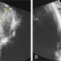
Ultrasound (US) is the primary modality for prenatal identification of triploidy. Before its widespread use, if analysis was not performed on aborted tissue, the diagnosis would usually go undetected. Triploidy should be suspected in any pregnancy with IUGR and fetal anomalies. The placenta usually demonstrates cystic changes in the type I phenotype ( Fig. 148.2 ), and is more commonly small and noncystic in the type II phenotype.

The diagnosis may be suspected in the first trimester by small crown-rump length, increased nuchal translucency, increased head-to-body ratio, oligohydramnios, and/or placental abnormalities. One study found increased nuchal translucency in type I, and increased head-to-body ratio in type II triploidy. From 2008–2014, one group identified 25 cases of triploidy (2 : 10,000 pregnancies) with transvaginal US at 12–16 weeks. Four of these manifested molar placental changes. Of the other 21, all were IUGR, and most (n = 20) had oligohydramnios and intracranial abnormalities (abnormal posterior fossa or enlargement of the fourth ventricle). Furthermore, 81% demonstrated absent gallbladder, 70% cardiac anomalies, and >50% renal anomalies and/or clenched hands.
In the second and third trimesters, major fetal malformations can more readily be seen ( Fig. 148.3 ). IUGR (most commonly with abdominal circumference lag) and oligohydramnios are present in most fetuses. US investigation of anomalies is often difficult because of oligohydramnios. Reported fetal anomalies are listed in Table 148.1 .


