Abstract
Trisomy 13 (Patau syndrome) is the third most common autosomal trisomy in newborns. It results from an extra chromosome 13 secondary to nondisjunction or translocation. In the United States, most cases of trisomy 13 are detected prenatally, either by genetic screening or ultrasound. Greater than 90% of fetuses with trisomy 13 have findings detected on prenatal ultrasound. The most common abnormalities visualized are cardiac abnormalities, holoprosencephaly, omphalocele, and cleft lip/palate. Fetuses with trisomy 13 have a 50% risk of intrauterine fetal demise after 12 weeks of gestation. Of live-born infants, most will not survive the immediate newborn period. Those that survive have severe physical and neurocognitive deficits.
Keywords
holoprosencephaly, microcephaly, cleft lip, cardiac defect
Introduction
Trisomy 13, also known as Patau syndrome, is one of the three most common trisomy syndromes. It is often diagnosed prenatally by the identification of one or more congenital abnormalities seen on ultrasound (US). Trisomy 13 is associated with severe physical and mental disabilities in addition to poor long-term survival rates in live-born infants.
Disorder
Definition
First genetically described by Patau et al. in 1960, trisomy 13 comprises a group of congenital abnormalities resulting from an extra chromosome number 13. The syndrome can also result from a translocation of chromosome 13.
Prevalence and Epidemiology
Trisomy 13 is estimated to occur in approximately 0.5–1.0 : 10,000 live births. The prevalence of trisomy 13 does not differ among races, and it occurs equally in both genders. However, a larger proportion of female fetuses survive to birth. Of live-born trisomy 13 cases, 5% are mosaic.
Etiology and Pathophysiology
Trisomy 13 results from an extra chromosome 13 secondary to nondisjunction or translocation. Approximately 75% of cases of trisomy 13 are due to nondisjunction. Abnormal ventral induction by the prechordal mesoderm of the prosencephalon is thought to be embryologic disturbance resulting in the brain and midfacial findings in trisomy 13. The mechanism by which the presence of an extra chromosome 13 leads to multiple anomalies is unknown.
Manifestations of Disease
Clinical Presentation
Because of routine prenatal US and screening in developed countries, most cases of trisomy 13 are identified in the prenatal period. Approximately half of pregnancies with trisomy 13 result in miscarriage or stillbirth between 12 weeks’ gestation and term.
Physical findings in live-born infants with trisomy 13 include holoprosencephaly, microphthalmia, cleft lip/palate, microcephaly, scalp defects, polydactyly, capillary hemangiomas, colobomas of the iris, and umbilical hernias. Most infants born with trisomy 13 die during the perinatal period. Severe mental and physical disability is seen in these infants. Isolated cases of survival into adulthood have been reported. One case series from Canada showed that surgical intervention can prolong survival, with approximately 70% surviving for 1 year after the first surgery, although these patients were likely to have been carefully selected due to the controversies surrounding surgical intervention in these cases.
Imaging Technique and Findings
Ultrasound.
US is the primary imaging modality for suspected cases of trisomy 13. One study showed that 95% of fetuses with trisomy 13 were detected on first-trimester or second-trimester US. Another study showed that all fetuses with trisomy 13 had one or more abnormalities or abnormal growth on US. In another study of 33 fetuses with trisomy 13, greater than 90% of affected fetuses had US findings suggestive of trisomy 13.
In the last decade, first-trimester combined screening and cell-free fetal DNA screening have emerged as the leading methods of diagnosis of trisomies, including trisomy 13. US findings include increased nuchal translucency above the 95th percentile for crown-rump length ( Fig. 149.1 ), decreased crown-rump length, congenital heart defects, holoprosencephaly, and omphalocele.

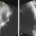
In the second trimester, the most common major abnormalities are congenital heart defects and central nervous system lesions (e.g., holoprosencephaly). Cardiac abnormalities are present in more than 50% of fetuses and include ventricular septal defects, hypoplastic left ventricle, and double-outlet right ventricle. Other findings include cleft lip/palate ( Fig. 149.2 ), hypotelorism, facial abnormalities, and postaxial polydactyly ( Fig. 149.3 ). Echogenic or polycystic kidneys and omphalocele ( Fig. 149.4 ) may also be seen. The finding of omphalocele increases the risk of trisomy 13 or trisomy 18 by 340-fold. Fetal growth restriction (less than the fifth percentile) in the second trimester is found in at least one-half of fetuses with trisomy 13.


