11
MODEL TUMOR SYSTEMS AND PREDICTIVE ASSAYS
C. MARC LEYRER, CAMILLE BERRIOCHOA, AND EHSAN H. BALAGAMWALA
Question 1
What are the five techniques to assay the response of solid tumors to treatment?
Question 3
What are the two methods used to measure tumor growth?
Question 1 What are the five techniques to assay the response of solid tumors to treatment?
Answer 1
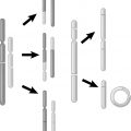
a. Tumor growth measurements.
b. Tumor cure assay.
c. In vivo dilution assay technique to determine tumor cell survival.
d. Lung colony assay to determine tumor cell survival.
e. In vivo treatment followed by in vitro assay for tumor cell survival.
Hall EJ, Giaccia AJ. Model tumor systems. In: Hall EJ, Giaccia AJ, eds. Radiobiology for the Radiologist. 7th ed. Philadelphia, PA: Lippincott Williams & Wilkins; 2012:356–371.
Question 3 What are the two methods used to measure tumor growth?
Answer 3
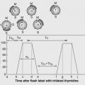
a. Using growth delay, which follows the time required for a tumor to return to the original size prior to radiation.
b. Evaluating the time required for a tumor to grow to a specified size after radiation therapy (RT), which is better utilized in tumors that do not respond to radiation.
Hall EJ, Giaccia AJ. Model tumor systems. In: Hall EJ, Giaccia AJ, eds. Radiobiology for the Radiologist. 7th ed. Philadelphia, PA: Lippincott Williams & Wilkins; 2012:356–371.
Question 4
What are the different types of programmed cell death?
Question 6
How long does it take for cells to show signs of death by apoptosis?
Question 4 What are the different types of programmed cell death?
Answer 4
a. Apoptosis—regulated cell death, which can occur in multicellular organisms and leads to characteristic morphological changes in the cell. This includes rounding up of the cell, plasma membrane blebbing, cell shrinkage, nuclear/chromosomal fragmentation, and chromatin condensation.
b. Autophagy—regulated cell death without chromatin condensation but instead by autophagic vacuolization of the cytoplasm containing degenerating organelles or cytosol.
c. Cornification—a specific type of programmed cell death occurring exclusively in the epidermis that is responsible for producing the dead keratinocytes necessary for the cornified skin layer. This process is often called keratinization.
d. Necrosis—cell death due to external factors and/or unfavorable conditions such as trauma, infection, and/or environmental changes.
e. Mitotic death—occurs during or shortly after a dysregulated/failed mitosis.
Kroemer G, Galluzzi L, Vandenabeele P, et al. Classification of cell death: recommendations of the Nomenclature Committee on Cell Death 2009. Cell Death Differ. 2009;16(1):3–11.
Question 6 How long does it take for cells to show signs of death by apoptosis?
Answer 6
Signs of cell death by apoptosis can occur as soon as 3 to 5 hours after irradiation.
Hall EJ, Giaccia AJ. Model tumor systems. In: Hall EJ, Giaccia AJ, eds. Radiobiology for the Radiologist. 7th ed. Philadelphia, PA: Lippincott Williams & Wilkins; 2012:356–371.
Question 7
What is cellular senescence?
Question 9
What is a tumor control/cure assay?
Question 7 What is cellular senescence?
Answer 7
Cellular senescence is a cellular state where the cell remains metabolically active but has permanently lost the ability to divide. This state can be initiated by many factors including telomere shortening, DNA damage, chromatin changes, and activation of cellular oncogenes.
Campis J, d’Adda di Fagagna F. Cellular senescence: when bad things happen to good cells. Nat Rev Mol Cell Biol. 2007;8(9):729–740.
Related posts:
 of CancerKailin Yang and Jennifer S. Yu
Influencing Tumor Control and ComplicationsDaesung Lee
Radiation Therapy ModalitiesSusan Kost and Andrew Godley
of CancerKailin Yang and Jennifer S. Yu
Influencing Tumor Control and ComplicationsDaesung Lee
Radiation Therapy ModalitiesSusan Kost and Andrew Godley
 Chromosome and Chromatid Damage, Repair, and MeasurementEhsan H. Balagamwala, C. Marc Leyrer, Jeffrey A. Kittel, and Alex Almasan
 and Risks in Nonradiation Oncology SubspecialtiesFrank Dong, Kevin Wunderle, and Salim Balik
 KineticsMatthew C. Ward and Richard Blake Ross
Stay updated, free articles. Join our Telegram channel

Full access? Get Clinical Tree


