Fig. 1
Metastatic renal cell carcinoma. (a) Surgical specimen: gross appearance of metastatic renal cell carcinoma appearing as a well-circumscribed yellow–orange to red–brown mass. (b) Macrosection: two nodules of metastatic renal cell carcinoma with hemorrhaged cystic degeneration
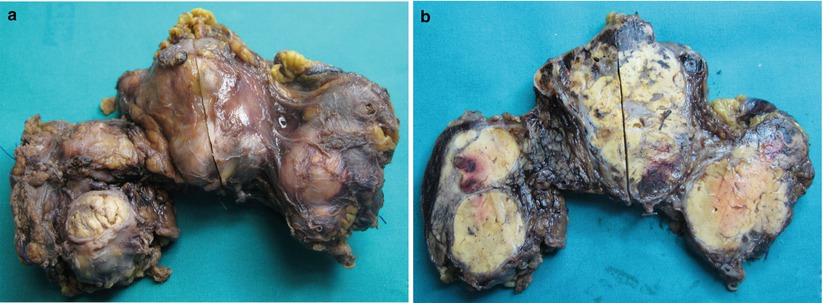
Fig. 2
Metastatic renal cell carcinoma. (a, b) Surgical specimen: multiple pancreatic lesions bulging the profiles of the gland (a). At cut section (b), multiple masses are clearly visible
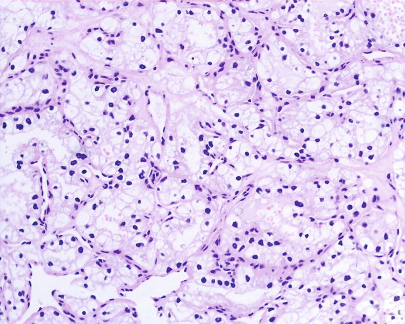
Fig. 3
Metastatic renal cell carcinoma. Histopayhology: sheets, small nests, and cords of clear cell, separated by rich sinusoidal vascular network
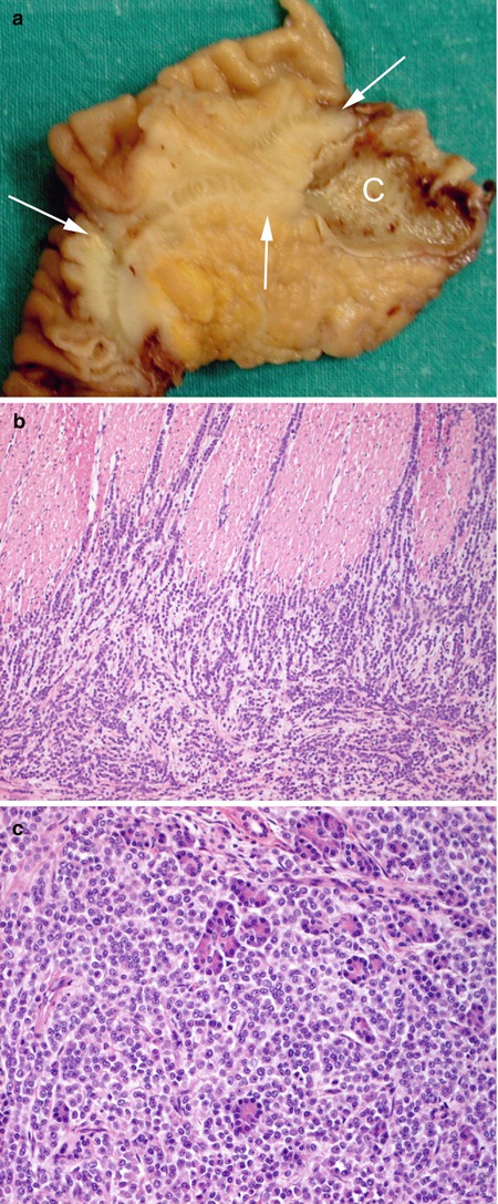
Fig. 4
Metastases of breast cancer. (a) Surgical specimen (pancreaticoduodenectomy): gross appearance of breast metastases with the neoplasm (arrows in a) mimics pancreatic adenocarcinoma with duodenal wall and common bile duct (C in a) infiltration. (b, c) Histopathology: the neoplastic cells involve the duodenal wall (b) and pancreatic lobules, replacing normal acinar tissue (c)
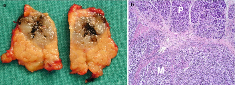
Fig. 5
Metastatic melanoma. (a) Surgical specimen (intermediate pancreatectomy): solid mass with black to brown area reflecting the presence of melanin pigment. (b) Histopathology: pancreas (P) and melanoma (M)
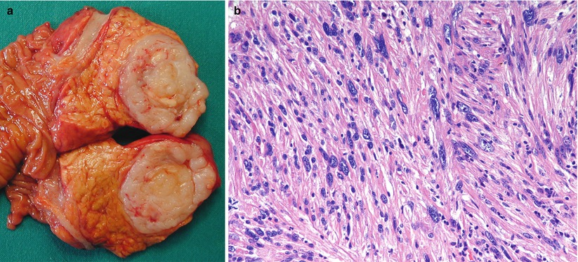
Fig. 6
Metastatic leiomyosarcoma. (a) Surgical specimen (pancreaticoduodenectomy): gross appearance of mesenchymal tumor with the mass appearing composed by soft tissue. (b) Histopathology: the tumor exhibits high cellularity, and it is composed of spindle cells with atypia

Fig. 7
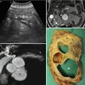
Hypervascular mets: metastatic renal cell carcinoma. (a) Ultrasound: single mass in the pancreatic head is visible, appearing solid and hypoechoic (arrow). The Wirsung duct (W) is dilated. (b, c) CT: the pancreatic head mass is confirmed and appears hypervascular (arrow in b). More than one lesion are visible; small hypervascular nodule (arrow in c) is in fact also detected. The Wirsung duct (W in c) is dilated
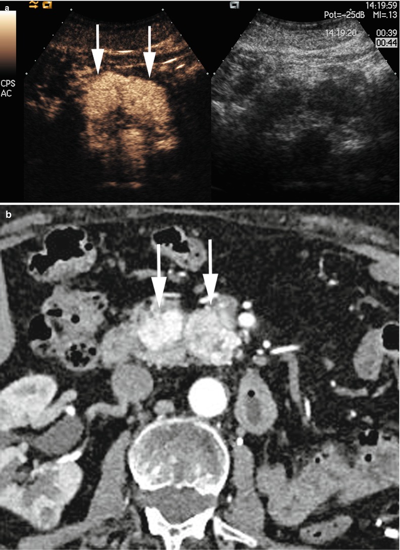
Fig. 8
Hypervascular mets: metastatic renal cell carcinoma. (a) CEUS: double hypervascular masses (arrows) in the uncinate process of the pancreas. (b) CT: double hypervascular masses (arrows) in the uncinate process of the pancreas
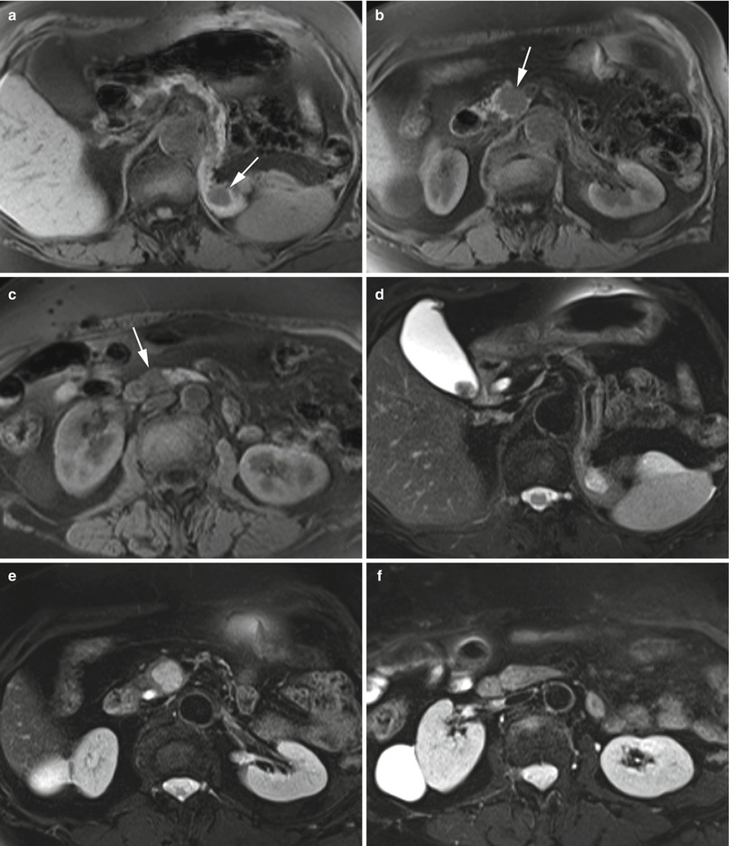

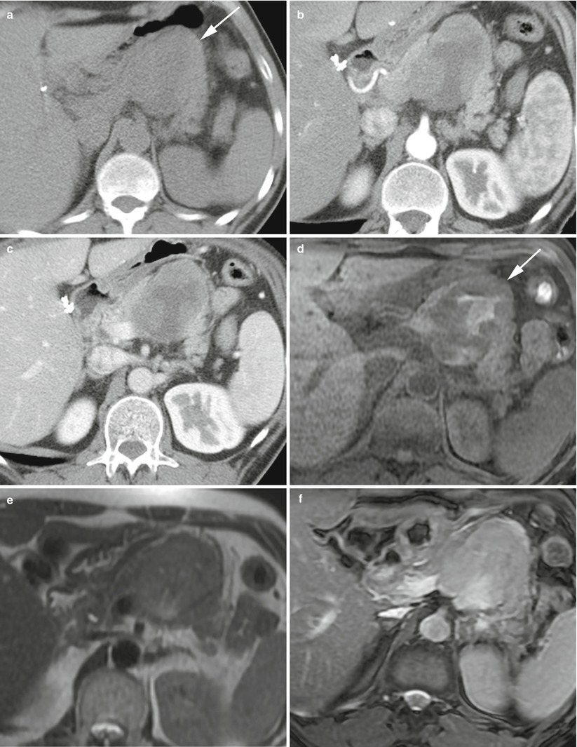
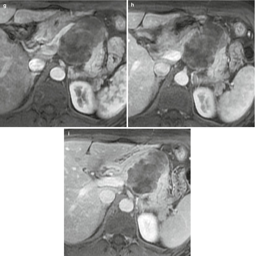
Fig. 9
Hypervascular mets: metastatic renal cell carcinoma. (a–l) MR study: on T1-weighted images (a–c), three hypointense nodules are visible; one in the pancreatic tail (arrow in a), one in the pancreatic head (arrow in b), and one very small in the caudal paraduodenal portion of the pancreatic head (arrow in c). On T2-weighted images (d–f), the lesions are visible as hyperintense, except the smallest in the caudal paraduodenal portion of the pancreatic head (f). On DWI, all the lesions show diffusion restriction at high b-value, appearing hyperintense (arrow in g–i). In the dynamic phase, the lesions are hypervascular with good conspicuity (arrow in j and k), except for the smallest one in the caudal paraduodenal portion of the pancreatic head (l)
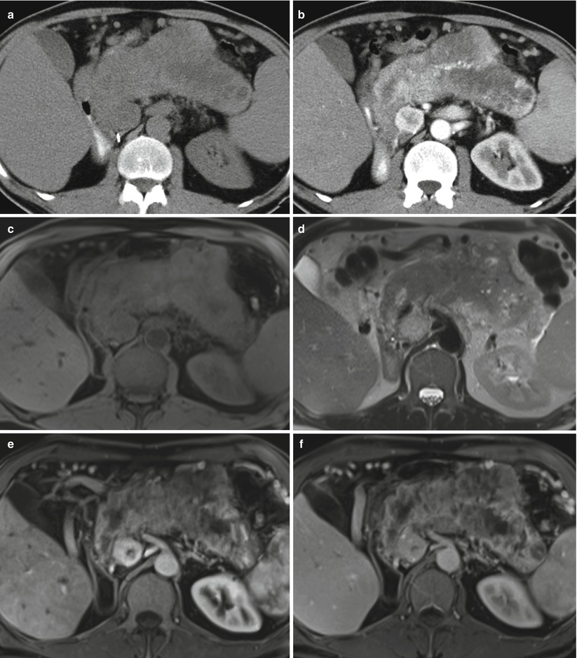
Fig. 10
Hypervascular mets: metastatic renal cell carcinoma. (a, b) CT study: in the baseline scan (a), enlargement of the pancreatic gland is immediately visible. In the pancreatic phase (b), a huge mass, inhomogeneously hypervascular, occupies the entire gland; moreover, another hypervascular nodule is visible in the uncinate process of the pancreas. (c–f) MR study: on T1-weighted images (c), the mass is inhomogeneously hypointense. On T2-weighted images (d), the mass is inhomogeneously hyperintense while the nodule visible in the uncinate process of the pancreas is homogeneously hyperintense. In the dynamic phases, pancreatic (e) and venous (f), the mass is inhomogeneously hypervascular with intralesional necrotic areas, and the nodule visible in the uncinate process of the pancreas is hypervascular with small intralesional central necrosis
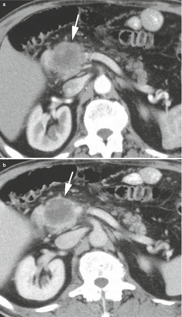
Fig. 11
Hypovascular met: metastatic lung cancer. (a, b) CT study: in the pancreatic (a) and venous (b) phases, a hypovascular mass (arrow in a and b) in the pancreatic head is visible
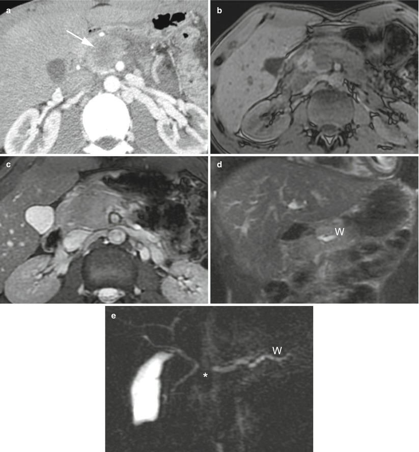
Fig. 12
Hypovascular met: metastatic rhabdomyosarcoma. (a) CT study: hypovascular mass (arrow) in the pancreatic head. (b–d) MR study: on T1-weighted images (b), the mass is hypointense. On T2-weighted images (c, d), the mass is inhomogeneously hyperintense. The Wirsung duct (W in d) is upstream-dilated. On MRCP (e), the mass stops (asterisk) the Wirsung duct (W in e) upstream-dilated