Etiology
The precise cause of gallbladder carcinoma is unknown, but cholelithiasis and pancreaticobiliary malformations are major risk factors. Gallstones and reflux of pancreaticobiliary enzymes are thought to result in chronic repetitive inflammation of the gallbladder mucosa that, over time, may undergo malignant transformation into invasive carcinoma.
Prevalence and Epidemiology
Gallbladder carcinoma is the most common primary biliary tract cancer and sixth most common gastrointestinal malignancy. It typically affects older patients (>65 years), with a female and Native American/Hispanic predominance. Associated conditions include chronic cholecystolithiasis; large (>3 cm) or cholesterol-type gallstones; gallbladder wall calcification (“porcelain gallbladder”); large (>1 cm) gallbladder polyps; obesity; anomalous pancreaticobiliary junctions and choledochal cystic disorders; chronic cholangitic infection by Salmonella or Helicobacter; inflammatory bowel disease and familial adenomatous polyposis; use of tobacco, estrogens, isoniazid, or methyldopa; and occupational exposures to rubber, petroleum, heavy metals, and radon.
Clinical Presentation
Signs and symptoms are nonspecific. In early stages, symptoms can mimic those of cholelithiasis or cholecystitis with right upper quadrant abdominal pain. Advanced disease can lead to significant biliary obstruction with resulting weight loss, hepatomegaly, jaundice, ascites, and less frequently bowel obstruction.
Most cases are advanced stage at diagnosis or less commonly incidentally discovered postoperatively. Poor prognostic factors include tumor invasion and metastatic spread at the time of diagnosis. When tumors are detected early, surgical resection can be curative.
On physical examination, classic signs include a painless enlarged gallbladder in the right upper quadrant (Courvoisier’s sign), periumbilical (Sister Mary Joseph node), or left supraclavicular (Virchow’s node) lymphadenopathy, and pelvic seeding palpable on digital rectal examination (Blumer’s shelf).
Pathophysiology
The gallbladder is a saccular organ located between the right and left hemilivers, just below segments IV and V. Sixty percent of tumors occur in the fundus, 30% in the body, and 10% in the neck ( Figure 53-1 ). Cancers disseminate via lymphatic spread to the porta hepatis, peripancreatic, and retroperitoneal nodes and via hematogenous spread to the lungs, liver, and bones. Direct local spread can lead to intraperitoneal seeding or infiltration of adjacent tissues. The American Joint Committee on Cancer Staging Tumor, Node, Metastasis (TNM) classification system is most commonly used for staging ( Figure 53-2 ).

Pathology
The gallbladder has three microscopic layers: mucosa, muscularis, and adventitia, or serosa. Serosa is found on the free surface of the gallbladder, and a layer of adventitia is present at liver interfaces. The muscularis layer consists of contractile smooth muscle, surrounded by a perimuscular connective tissue sheath. The mucosa consists of lamina propria and simple columnar epithelium lining the gallbladder lumen. There is no muscularis mucosa or submucosa separating the muscularis and mucosa layers, leading to easier spread of tumor into the adjacent liver and peritoneum.
Ninety percent of gallbladder cancers are adenocarcinomas that originate from glandular cells in the gallbladder lining. Subtypes include papillary and mucinous tumors. The papillary type is least aggressive and has the best prognosis. Other histologic types include squamous cell, adenosquamous, small cell, neuroendocrine, and sarcomatoid. Occasionally, metastases from melanoma, renal, and breast carcinoma can involve the gallbladder. Histologic grade also affects outcome, with poorly differentiated tumors portending a worse prognosis.
Benign tumors of the gallbladder include adenomas, villous papillomas, paragangliomas, and granular cell tumors. Adenomas are uncommon lesions that result from chronic inflammation and/or cholelithiasis.
Imaging
Gallbladder carcinoma manifests either as an intraluminal polypoid or sessile mass, gallbladder wall thickening, or a large extraluminal mass invading surrounding organs. Most tumors are seen in the fundus or body, with fewer in the neck or cystic duct. Irregular mural thickening and gallbladder calculi may also be present.
Radiography
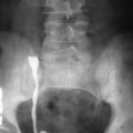
Plain radiography has limited value in gallbladder carcinoma. Abdominal films may show porcelain gallbladder, calcified gallstones, vague or punctate tumor calcification, and occasionally pneumobilia from a gallbladder-enteric fistula ( Figure 53-3 ). Invasive procedures such as endoscopic retrograde cholangiography (ERCP) or percutaneous transhepatic cholangiography (PTC) can show bile duct narrowing and/or nonvisualization of the gallbladder secondary to obstruction ( Figure 53-4 ). A malignant-appearing stricture of the midportion of the common bile duct (CBD) should raise suspicion for a gallbladder carcinoma.
Computed Tomography
Computed tomography (CT) can detect gallbladder masses and wall thickening. Tumors are visualized as hypodense masses that enhance heterogeneously, occasionally filling the entire lumen (the “jam-packed gallbladder”). Focal or diffuse wall thickening also can be seen, often with abnormally avid or persistent enhancement. Porcelain gallbladder, calcified gallstones, and/or biliary dilatation also may be present. Advanced tumors may manifest with hepatoduodenal ligament lymphadenopathy and invasion of the liver, bile ducts, duodenum, stomach, pancreas, and/or kidneys. Metastasis may be noted in distant organs ( Figure 53-5 ).
Magnetic Resonance Imaging
Findings on magnetic resonance imaging (MRI) are similar to those of CT, with superior assessment of local disease extension and tumor composition because of better soft tissue contrast. Tumors generally appear hyperintense on T2-weighted and isointense to hypointense on T1-weighted images. When gadolinium is administered, tumors enhance heterogeneously and peripherally ( Figure 53-6 ). Magnetic resonance cholangiopancreatography (MRCP) provides superior visualization of the biliary system for assessing obstruction ( Figure 53-7 ). High-resolution three-dimensional T1-weighted gradient echo postcontrast MR images delineate vascular detail for surgical planning. More recently, hepatobiliary contrast agents have been advocated to improve detection of liver invasive disease.

Stay updated, free articles. Join our Telegram channel

Full access? Get Clinical Tree


