Chapter Outline
Ultrasound is an ideal modality for examination of the solid abdominal viscera. It is relatively inexpensive and portable and does not require the use of potentially harmful ionizing radiation or contrast agents. It enables real-time examination and can be used in the emergency department and by the bedside in the wards. Continual improvements in ultrasound technology and computing during the six decades since medical ultrasound was first developed have ensured that it remains the first-choice modality for many applications within the abdomen. Ultrasound has several advantages over computed tomography (CT) and magnetic resonance imaging (MRI), including cost (and hence ease of access), real-time imaging, portability, lack of ionizing radiation, and absence of contraindications, although the cross-sectional modalities are often complementary. Further advances in ultrasound technology will ensure that it retains a central role in abdominal imaging for the foreseeable future.
The first use of medical ultrasound was documented in Austria in 1942 by Karl Dussik, who imaged the ventricles of the brain and brain tumors. Another pioneer of medical ultrasound, Ian Donald in Glasgow, built on innovations in the fields of sonar, radar, and ultrasonic flaw detection in metals to image uterine fibroids and ovarian cysts. He also published a paper entitled “The Investigation of Abdominal Masses by Pulsed Ultrasound” in 1958. Several technologic innovations were necessary to drive ultrasound forward as the commonplace medical imaging technique we know today. The first static arm commercial medical scanners were marketed in the 1960s, but the technique was slow and the machines were cumbersome. Real-time scanners soon replaced static arm scanners. These early scanners used the A-mode technique, in which the amplitude of returning echoes along a single line was displayed on an oscilloscope screen. Later B-mode compound scan images were composed of only black and white. Gray-scale imaging improved interpretation and is now the standard for abdominal imaging. The development of the transistor and later the integrated circuit improved signal generation and amplification. All modern machines now employ digital signal processing techniques, and improvements in the design and manufacture of probes have allowed the development of compact, robust, and versatile probes for a variety of applications.
The real-time nature of imaging with ultrasound lends itself particularly well to problem solving. Movement with respiration can be examined, tubular structures can be followed, and structures can be examined in different planes. Ultrasound can aid interventional procedures, such as fine-needle aspiration, core biopsy, and drain placement, and it is the modality of choice for guiding interventional procedures within the abdominal solid organs, although on occasion it may be necessary to supplement it with fluoroscopy, CT, or MRI. However, the real-time nature of ultrasound requires competence on the part of the ultrasonographer or sonologist performing the examination. Although the examination is performed in real time with up to 60 frames per second, only a fraction of these are saved as representative images for future review. Thus the experience of the operators is important because they will decide on the images to store. Picture archiving and storage (PACS) systems have enabled more still images to be stored without incurring the cost of printing film and may also allow the storage of cine loops to show dynamic processes.
Ultrasound enables blood flow to be easily demonstrated without the need for nephrotoxic contrast agents, and neither the patient nor the operator is exposed to harmful ionizing radiation. The ability of ultrasound to differentiate between solid and cystic structures is a particular strength.
Basic Physics
Medical ultrasound uses short pulses of sound waves that are transmitted and received by the transducer ( Fig. 66-1 ). A coupling gel is used to facilitate the transmission of sound waves to and from the probe. This matches the acoustic impedance of the probe to that of tissue and eliminates air gaps between the probe and the skin. The basic element of most current transducers is a piezoelectric crystal that emits a sound wave when a voltage is applied to it (transmission) and generates a voltage when the reflected sound wave returns to it (reception). The voltages produced by each crystal on receiving reflected sound waves are converted into digital signals by an analog to digital signal converter and processed by the ultrasound machine to form the image. Future transducer designs may use alternative methods of transducer construction, but the principles of sound wave generation and reception will remain the same. The probe contains multiple crystals arranged in a strip or a grid. There are typically 128 individual crystals within a transducer array, and they can be independently activated; this allows the ultrasound beam to be electronically steered and focused. Earlier designs used mechanical devices to steer and focus the beam, but these have largely been superseded by electronic controls. The best resolution of an image is within the focal zone, and focusing allows the focal zone to be varied according to the area of interest being examined. The beams of array transducers can be focused on both transmit and receive. Some transducers allow multiple focal zones to be selected to increase the effective focal zone, but this will reduce the frame rate or temporal resolution of the scan.

Several factors have to be taken into account in considering the most appropriate choice of transducer for a given application. Modern transducers suitable for abdominal imaging include linear arrays, curved linear arrays, and sector scanners. Linear arrays (typically 7-12 MHz) provide excellent resolution at the expense of penetration and field of view. They are ideal for superficial lesions or for examining the surface of abdominal viscera, such as the liver ( Fig. 66-2 ). The field of view can be extended by the use of trapezoidal imaging ( Fig. 66-3 ) or compound (or extended field of view) imaging, in which the probe is moved across a structure and the ultrasound machine “stitches together” the images to produce a panoramic image. This is useful for large superficial lesions, such as soft tissue tumors ( Fig. 66-4 ). Curved linear (or curvilinear) arrays are less effective for the examination of superficial structures but provide better penetration (because of their lower frequencies) and a wider field of view. Sector scanners have a limited role in routine abdominal ultrasound but do provide a wide field of view with a small footprint, which can be useful when access is limited, for example, in examining the pediatric abdomen. A compromise must be reached between penetration and resolution, which are inversely proportional. Raising the frequency of the transmitted ultrasound pulse will improve resolution because of shortening of the wavelength, but it will also decrease the penetration as high frequencies are attenuated more than low frequencies are (attenuation of the sound wave is proportional to frequency). Many modern transducers allow the operator to vary the transmitted frequency within a certain range without having to change probes, for example, from 1 to 5 MHz for a typical curvilinear probe. Most modern ultrasound machines will select the transmitted frequency on the basis of the user’s selection of presets, optimizing the image for detail or resolution (high frequency) or penetration (low frequency).


Sound waves interact with the tissues of the body in three main ways: reflection, absorption, and scatter. The terminology used to describe ultrasound findings is a description of the brightness of the structure; the more sound energy reflected back to the transducer, the brighter the object on the image. Objects are described as hypoechoic (darker than adjacent structures), isoechoic (the same brightness as adjacent structures), or hyperechoic (brighter than adjacent structures). Anechoic structures appear black and indicate fluid. Internal echoes may be caused by debris, septations, or artifacts. Often the liver is chosen as a reference in the abdomen, but care must be taken to ensure that the reference organ is of normal reflectivity and not abnormal due to fatty infiltration, for example. The amount of energy reflected back to the transducer depends on several factors. The simplest form of reflection occurs when sound waves encounter a flat interface perpendicular to the beam with a large disparity in the velocity of sound between the adjacent tissues; this is termed a specular reflector and will appear as a well-defined bright line ( Fig. 66-5 ). Smaller interfaces (0.1-1 mm in diameter) may cause scattering in all directions. Only a small fraction of the transmitted energy is returned to the probe, but when multiple such structures are present, the interference pattern between echoes produces a visible texture. This process is responsible for the ultrasound appearance of liver, spleen, and kidney parenchyma.

Sound waves propagate through tissues at different velocities, and when there is an interface between different tissues, the sound may be reflected or refracted (just as light may be reflected or refracted by a prism of glass). The greater the difference in velocity between the adjacent tissues, the greater proportion is reflected (if the incident angle of the beam is large) or the more the beam is refracted (if the incident angle of the beam is smaller) ( Fig. 66-6 ).

Recent Developments
Ultrasound machines continue to rapidly evolve with the introduction of new technology. Although the principles of physics remain the same, manufacturers have developed new methods of probe construction, ultrasound generation, and postprocessing algorithms. Often these are assigned brand-specific trademarked titles that may be rather opaque to the user.
Many ultrasound machines now allow the user to select a real-time compound imaging mode, whereby the area to be examined is insonated from several different directions by electronic beam steering for transmission and reception. The displayed image is an average of the images derived from different insonation angles. This has been shown to improve overall image quality, to increase lesion conspicuity, and to reduce artifact. However, it does reduce the frame rate and can lead to image blurring if the transducer or the subject is moving.
A recent development that is now standard on most machines is tissue harmonic imaging. “Conventional” imaging without harmonics is known as fundamental imaging because only the fundamental transmitted frequency is used for transmission and reception. Tissue harmonic imaging was originally proposed for use with an ultrasound contrast agent, but it has been shown to improve visualization of abdominal structures without the use of contrast agents. The shape of the transmitted waveform changes as a sound wave is propagated through tissue, particularly in the focal zone, where the intensity is high. Tissues resist compression more than expansion. This is termed nonlinear behavior. The waveform of the reflected pulse is altered and contains higher frequencies than the transmitted pulse. The higher frequencies are multiples of the transmitted fundamental frequency, called harmonic frequencies. At present, it is usually the second harmonic (twice the fundamental frequency) that is used for imaging, but higher multiples may also be used. The fundamental frequencies are electronically filtered out after reception, leaving only the harmonic frequencies. The harmonic frequencies travel through the tissues only once on the return to the probe and therefore are attenuated half as much as an identical frequency transmitted and received by the probe. There is less scatter, particularly in obese patients, in whom subcutaneous fat and superficial muscle layers cause scatter and attenuation of the fundamental frequencies, which may cause a haze of echoes, degrading the image on fundamental imaging. Artifacts such as side and grating lobe artifacts that degrade fundamental images are also reduced because the weak side and grating lobes are not powerful enough to produce harmonic frequencies. The images are clearer, and tissue harmonic imaging may improve diagnostic ability in challenging patients. Phase inversion imaging also exploits the nonlinear behavior of tissues but employs a different technique from frequency-based tissue harmonic imaging. Two pulses are sent with 180-degree phase difference. If the pulse is reflected in a linear fashion, the reflected waves will cancel each other out. If the waves are reflected in a nonlinear fashion, the waves will not cancel out and are used to form the image. Phase inversion harmonic imaging reduces the frame rate because of the need to transmit twice as many pulses, but the spatial resolution is better than that of frequency-based tissue harmonic imaging.
Three-dimensional and four-dimensional ultrasound applications are established in obstetric ultrasound, but their value is yet to be proved in abdominal imaging. Improvements in processing and probe technology have made real-time three-dimensional volume rendered and multiplanar reconstruction imaging possible. An acquisition of a volume of data in a fashion akin to helical CT allows retrospective reconstruction of multiplanar reconstruction views and surface rendered images.
Doppler Ultrasound
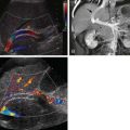
The examination of blood flow with Doppler techniques is a valuable addition to real-time gray-scale abdominal ultrasound. The wavelengths of a transmitted sound pulse and the received pulse differ when sound waves are reflected from a moving surface. The wavelength shortens when the reflector is moving toward the transducer and lengthens when it is moving away. The change in wavelength is accompanied by a change in frequency and phase and is termed the Doppler effect. The Doppler frequency (or phase) shift is measured, and the velocity and direction of blood flow can be calculated. One method of displaying the information is as color flow, in which a color map of flow is superimposed on a gray-scale real-time image. Convention dictates that blood flow toward the transducer is red and blood flow away from the transducer is blue, although the operator may invert this. Color flow Doppler is used in the abdomen to examine the arterial tree, the portal venous system, and the systemic veins ( Fig. 66-7 ). It may also be used to examine vascularity within the solid organs, for example, within or surrounding a mass lesion. The box displaying the color information can be resized and moved to include different parts of the image. A compromise between the color box size and the refresh rate of the image must be reached as an increased color box size will slow down the refresh rate of the image.

Power Doppler is a similar technique, but the total power of the reflected Doppler signal is displayed. There are no velocity data and therefore no indication of the direction of flow, but there are some advantages of power Doppler over color flow Doppler in certain situations. Power Doppler is more sensitive to slow flow and, unlike color Doppler, can display flow that is almost perpendicular to the beam. Background noise appears as brightly colored speckles in color Doppler that can be distracting, whereas in power Doppler the background noise is dark, allowing higher gains to be used. It is thus a useful technique when the demonstration of low flow is needed but the direction of flow is not important.
The waveform and the direction of flow in vessels may change in disease states, such as portal hypertension. Pulsed-wave Doppler displays the flow velocity as a waveform, and this is often combined with gray-scale imaging as duplex imaging ( Fig. 66-8 ). The flow is measured in a small sample volume that can be placed on the region of interest. The operator can adjust the position and the size of the sample volume. If quantitative measurement of flow is required, a correction has to be made as the Doppler beam is at an angle to the blood flow. The angle between the blood flow and the interrogating Doppler beam should be kept as small as possible; it is not possible to obtain a reliable Doppler velocity measurement if this angle exceeds 60 degrees. This quantitative data can be used to assess vascular resistance, for example, within a transplanted liver.

Ultrasound Contrast Media
Conventional ultrasound does have some limitations in the detection and characterization of liver lesions. Alternative cross-sectional imaging techniques, such as CT and MRI, have often been used to complement ultrasound to overcome these limitations. Ultrasound contrast agents have been developed to increase the sensitivity of ultrasound to small liver lesions and to improve the characterization when lesions are detected on the basis of their perfusion properties. The first-generation ultrasound contrast agents were based on air-filled bubbles and required high acoustic pressures (high mechanical index) to break the bubbles. Contrast enhancement was short-lived. Second-generation contrast agents are now available. These form small bubbles filled with gases other than air, such as sulfur hexafluoride, and allow real-time scanning at a low mechanical index depicting the different phases of blood flow, which is particularly useful in the liver. The appearances of a lesion in arterial, portal venous, and sinusoidal phases (15-35, 35-90, and 90-240 seconds, respectively) can be observed.
Most modern ultrasound machines include technology that will allow imaging with contrast agents, but an estimated 60% of ultrasound machines currently in use worldwide do not have the capability. The contrast agent is administered through an intravenous cannula as a rapid bolus followed by a saline flush. The area is then scanned continuously to depict the different vascular phases, and the images are often recorded as a cine clip to allow later playback. Repeated injections of contrast agent are possible, if necessary, as the agents are of low toxicity.
The detection of subcentimeter liver metastases is improved when contrast media are used. Contrast media can also be useful in differentiating benign from malignant tumors and characterizing the lesions. It has been suggested that a contrast agent should also be used in the assessment of abdominal trauma, in which it may be added to the FAST (focused assessment with sonography in trauma) protocol.
The use of ultrasound contrast agents remains highly variable, despite some encouraging clinical results. This is due to many factors, including workflow issues, availability of suitable ultrasound machines, and remuneration. It seems unlikely that they will become a method of choice for characterization of liver lesions, as some proponents suggest.
Ultrasound Artifacts
The sonologist should be familiar with common artifacts to minimize their effect on image quality and to avoid confusion with significant pathologic changes, although some of the processes described as artifacts can be helpful in interpreting images.
Several assumptions are made in forming an ultrasound image. It is assumed that the ultrasound beam travels in a straight line from the probe and in the body. The velocity of the sound wave is assumed to be constant on the path, and the received echoes are assumed to have traveled in a straight line back to the probe. If any of these assumptions are incorrect, an artifact will be produced.
If the ultrasound wave is propagated through a substance with reduced attenuation compared with the surrounding tissues, the region beyond it will appear brighter or more echogenic. This is due to the fact that a time gain compensation function is applied to the image, whereby the received echoes are amplified proportional to the time interval between pulse transmission and echo reception. The sound wave is attenuated as it passes through a structure, and the reflected echoes are also attenuated on their return to the transducer. If time gain compensation were not applied, deeper structures would appear unacceptably dark. If a structure, for example, a fluid-filled cyst, transmits sound waves effectively with little attenuation, the tissue beyond the cyst will appear brighter than equivalent adjacent tissue ( Fig. 66-9 ). This phenomenon is called acoustic enhancement, and it is an important feature of fluid-filled lesions. Conversely, if a structure attenuates more than the surrounding tissue, tissues beyond it will appear darker and lie within the acoustic shadow. This is typical of solid lesions ( Fig. 66-10 ). A very reflective surface, such as bone or air, will reflect the ultrasound beam and appear as a bright line with a very dark shadow beyond ( Fig. 66-11 ). The ability to differentiate between solid and cystic lesions is an important strength of ultrasound. Gas collections and gas within the bowel tend to produce “dirty” shadows, which are less clear than the shadows produced by a solid lesion such as a calculus.


Related posts:


Stay updated, free articles. Join our Telegram channel

Full access? Get Clinical Tree


