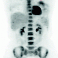
Fig. 7.1
The MIP image documents the presence of lymph node metastases. There are not secondary lesions in any other organs or systems
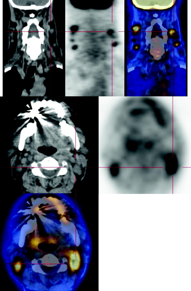
Fig. 7.2
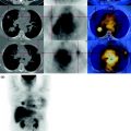
In correspondence with the lateral triangle of the neck, CT-PET shows multiple nodes bilaterally increased in size, some, measuring more than a centimeter, characterized by intense glucose metabolism
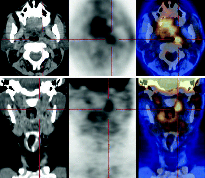
Fig. 7.3
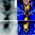 Laryngeal Squamous Carcinoma: Staging
 Sigmoid Adenocarcinoma: Post-Actinic Tardive Sacral Fracture
 Urothelial Carcinoma: Follow-Up After Surgery
 Lymphocytic Interstitial Pneumonia in Patient with History of Breast Cancer
 Metastatic Breast Carcinoma: Restaging After Neoadjuvant Chemotherapy
 Bone Metastases from Breast Cancer: Progression of Disease and Subsequent Response to Radiotherapy


CT-PET: presence of hypodense tissue in the left retropharyngeal region, with limited metabolism, therefore, to be attributed to post—actinic remodeling
Related posts:
Laryngeal Squamous Carcinoma: Staging
Sigmoid Adenocarcinoma: Post-Actinic Tardive Sacral Fracture
Urothelial Carcinoma: Follow-Up After Surgery
Lymphocytic Interstitial Pneumonia in Patient with History of Breast Cancer
Metastatic Breast Carcinoma: Restaging After Neoadjuvant Chemotherapy
Bone Metastases from Breast Cancer: Progression of Disease and Subsequent Response to Radiotherapy
Stay updated, free articles. Join our Telegram channel
Full access? Get Clinical Tree
