KEY FACTS
Terminology
- •
Also known as pelviureteric junction obstruction
- •
Obstruction of urine flow at level of UPJ
- •
Congenital or acquired
Imaging
- •
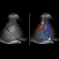
Marked hydronephrosis to level of UPJ without dilatation of ureter
- •
Renal pelvis disproportionately larger than calyces
- •
Color Doppler may detect crossing vessel
- •
Nuclear renal scan: Hydronephrosis with poor drainage, suggesting obstruction
- •
Ultrasound is 1st-line modality
- •
CTA or MRA for vascular evaluation prior to surgery
- •
Nuclear renal scan to determine relative renal function and confirm obstructive hydronephrosis
- •
Imaging recommendations
- ○
CT or MR to detect crossing vessel
- ○
Top Differential Diagnoses
- •
Multicystic dysplastic kidney
- •
Hydronephrosis of other etiology
- •
Extrarenal pelvis
- •
Pararenal cyst
Pathology
- •
Intrinsic causes: Abnormal peristalsis at UPJ, stone, clot, tumor, stenosis, scarring
- •
Extrinsic causes: Crossing vessels near UPJ
- •
Associated with renal ectopia and fusion anomalies
- •
Higher incidence in multicystic dysplastic and duplicated kidneys
Clinical Issues
- •
M > F, twice as common on left, 10-30% bilateral
- •
1 in 1,000-1,500 newborns
- •
Most common cause of antenatal and neonatal hydronephrosis, diagnosed by prenatal screening
- •
Symptoms include: Intermittent abdominal or flank pain, nausea, vomiting, failure to thrive, hematuria, renovascular hypertension (rare)
- •
Prognosis generally good, depends on degree of preserved renal function
- •
Treatment: Pyeloplasty (open, laparoscopic, or robotic-assisted laparoscopic)
Scanning Tips
- •
Use color Doppler for bladder jets and crossing vessels
 and calyces
  in a ureteropelvic junction obstruction. The ureter
  is not dilated.
 as well as the renal calyces
  . Note the severe cortical thinning
  .
.
 and the calyces
  .
.
Stay updated, free articles. Join our Telegram channel

Full access? Get Clinical Tree


