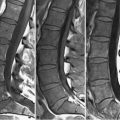
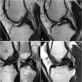
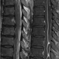
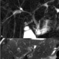
73 Urinary Bladder and Male Pelvis Urinary bladder carcinoma, a case of which is illustrated in Fig. 73.1, is best locally staged with MRI. (A) Sagittal FSE T2WI illustrate high SI urine, moderate SI bladder mucosa, low SI bladder musculature, and high SI perivesicular fat, the latter useful for delineating the bladder wall. A thickened wall (>5 mm) is a nonspecific finding seen in an underfilled bladder, acute cystitis, fibrosis, and infiltrative cancer. On (B) axial GRE T1WI low SI urine is surrounded by the moderate SI wall. T1WI readily detect superficial mucosal lesions as well as enlarged, low SI lymph nodes contained in perivesicular fat. Use of GRE T1WI, in addition to short acquisition times and adaptability to 3D acquisitions, allows distinction of nodes from high SI vascular structures. In Fig. 73.1A, an intermediate SI mass-like lesion interrupts the low SI of the muscular layer (white arrow); (B) T1WI demonstrates a moderate SI lesion involving the right bladder wall without infiltration of the surrounding fat. Carcinoma enhances earlier and more avidly than the normal wall. Delayed (>2 minutes) postcontrast image acquisition limits evaluation of the bladder wall due to urinary excretion of the administered gadolinium chelate. Cystitis enhances similarly, as opposed to the late enhancement seen with wall fibrosis. Bladder carcinoma is staged by the TNM (tumor, node, metastasis) system based on whether the tumor is superficial to the muscular layer (T1), invades the layer superficially (T2) or deeply (T2b), invades the perivesicular fat micro- (T3a) or macroscopically (T3b), or extends to adjacent organs (T4). An intact low SI muscular layer signifies a T1–T2 lesion on MRI, whereas T2b and T3a lesions (see Fig. 73.1
![]()
Stay updated, free articles. Join our Telegram channel

Full access? Get Clinical Tree


