Etiology
Urinary tract obstruction (UTO) is a syndrome that may be caused by a wide range of pathologic processes. It may vary in the following:
- •
Degree: May be partial or complete.
- •
Site: May be unilateral or bilateral and may occur at any level of the urinary tract from the calyces to the urethral meatus.
- •
Duration: May be acute or chronic.
- •
Demographics: Common causes vary among prenatal, neonatal, pediatric, young adult, and older adult patient groups and between males and females.
- •
Physiologic response: Can be compensated or uncompensated.
- •
Presence or absence of a morphologic stricture: Anatomic versus functional obstruction. Anatomic or mechanical obstruction is due to a fixed point of narrowing or an obstructing lesion. In functional obstruction, there is no demonstrable fixed narrowing, but, nonetheless, proximal pressures are raised (e.g., primary obstructive megaureter and some forms of pelviureteral junction obstruction).
Many of the causes of UTO are presented in Table 66-1 .
| Type of Obstruction | Kidney/Renal Pelvis | Ureter | Bladder | Urethra |
|---|---|---|---|---|
| Intraluminal | Staghorn calculus | Calculus Transitional cell carcinoma Sloughed papilla Blood clot | Calculus Transitional cell carcinoma | Posterior urethral valves |
| Intramural | Pelviureteral junction obstruction Infundibular stenosis | Stricture (e.g., postinfection, surgery or radiation therapy) Ureterocele Vesicoureteral reflux | Neuropathic bladder | Urethritis Stricture |
| Extramural | Retrocaval ureter Extrinsic tumor Retroperitoneal lymphadenopathy Retroperitoneal abscess Retroperitoneal fibrosis Inflammatory abdominal aortic aneurysm Large abdominal aortic aneurysm or iliac artery aneurysm Endometriosis Pregnancy | Benign prostatic hypertrophy Prostate carcinoma |
Prevalence, Epidemiology, and Definitions
UTO is a common clinical and uroradiologic diagnostic problem. The incidence of hydronephrosis in one autopsy series was 3.1%, with no difference in incidence between the sexes in patients younger than age 20 years. It is more common in women than men between 20 and 60 years of age (owing to obstetric and gynecologic causes) and more common in men older than 60 years (predominantly owing to benign prostatic hyperplasia). Hydronephrosis is the most common cause of an abdominal mass in neonates, and UTO is the most common cause of end-stage renal failure in children.
UTO has been defined as “a narrowing such that proximal pressure must be raised to transmit usual flow.” It is important to understand that this definition does not refer to dilation of the urinary tract. UTO may occur without dilation ( Box 66-1 ) and dilation may occur without UTO ( Box 66-2 ), creating potential causes of false-negative and false-positive findings in radiologic investigations of UTO.
- •
Retroperitoneal fibrosis
- •
Retroperitoneal tumor encasing ureters or renal pelvis, with loss of normal distensibility of collecting system
- •
Obstruction with urine extravasation, resulting in decompression of pelvicalyceal system
- •
Hyperacute urinary tract obstruction
- •
Vesicoureteral reflux
- •
Primary megaureter
- •
Previous obstruction
- •
Infection (pyelonephritis, peritonitis)
- •
High-flow states (diabetes insipidus, psychogenic polydipsia)
- •
Prune belly syndrome
- •
Congenital megacalyces
- •
Beckwith-Wiedemann syndrome
Other useful definitions include the following:
- •
Hydronephrosis (or pelvicaliectasis, pyelocaliectasis) refers to dilatation of the collecting system. This is often, but not always, due to UTO.
- •
Obstructive uropathy is synonymous with UTO. It describes the state in which there is increased resistance to urine flow.
- •
Obstructive nephropathy refers to renal damage caused by UTO. Over time, UTO results in medullary and cortical atrophy secondary to irreversible nephron loss.
Clinical Presentation
The presentation of UTO varies with the underlying cause. Acute UTO often manifests as pain, decreased urine output, and signs and symptoms of acute renal failure. Chronic UTO is often insidious. Patients may present with hypertension, irreversible chronic renal failure, recurrent urinary tract infections, or change in micturition. Upper tract obstruction may manifest with flank, back, or groin pain and lower tract obstruction with voiding dysfunction or suprapubic pain.
Pathophysiology
UTO may be caused by a pathologic process at any level of the urinary tract from the calyces to the urethral meatus. The resultant pathophysiology of UTO is complex. Collecting system pressure and renal blood flow (RBF) are important factors. In simple terms, UTO leads to increased collecting system pressure proximal to obstruction, which leads to the following:
- •
Initial transient renal vasodilation
- •
Increased RBF then vasoconstriction
- •
Increased resistance to RBF
- •
Decreased diastolic flow
- •
Ischemia
- •
Atrophy
In addition, UTO results in reduced urine concentration, reduced urine acidification, and abnormalities of electrolyte excretion. UTO causes urinary stasis, predisposing to infection and stones. If untreated, UTO causes progressive and eventually irreversible structural renal changes, including tubular atrophy, tubulointerstitial fibrosis, interstitial inflammation, and glomerular loss.
Recovery of renal function after relief of obstruction may be complete if obstruction is brief, but complete UTO lasting longer than 24 hours may cause irreversible loss of renal function. Increased age, lower baseline renal function, and high-grade and lengthy obstruction are factors associated with greater residual renal impairment.
Pathology
Macroscopically, the obstructed kidney may demonstrate mild to marked enlargement. There is progressive blunting of the apices of the medullary pyramids, which eventually become cupped. Variable parenchymal atrophy may be evident. In advanced obstruction, there may be complete obliteration of the pyramids, marked cortical thinning, and massive dilation of the collecting system.
Early renal microscopic changes include edema and tubular dilation. Increasing edema widens Bowman’s space, followed by thickening of the tubular basement membrane, then papillary necrosis, infiltration with inflammatory cells, and interstitial fibrosis. In the end stages, there is glomerular collapse, tubular atrophy, and connective tissue proliferation.
Imaging
When interpreting radiologic studies of the urinary tract, anatomic and functional factors should be considered and modalities that depict one without the other should be interpreted with caution.
Plain Radiography
Ninety percent of urinary calculi are radiopaque and therefore theoretically visible on plain radiographs. Factors limiting visualization include stone composition and size, patient habitus, extraurinary calcifications, and overlying bowel gas and content. Radiolucent calculi (pure urate or xanthine calculi, matrix stones) may not be detected, nor may noncalculous causes of obstruction.
Excretory Urography
Excretory urography (EUG), also referred to as intravenous pyelography (IVP), was previously the gold standard in the assessment of UTO. It provides anatomic and functional information.
Excretory Urography Findings of Acute Urinary Tract Obstruction
The principle EUG findings in acute UTO are as follows:
- •
Immediate nephrogram usually normal or mildly diminished (reflects normal RBF)
- •
Increasingly dense nephrogram over time
- •
Delayed excretion of contrast agent into the collecting system
- •
Variable dilation of collecting system proximal to point of obstruction
Other findings may include the following:
- •
Heterotopic excretion of contrast via the biliary system, leading to opacification of the gallbladder
- •
Urine or contrast extravasation occurs if renal pelvis pressure is sufficiently high
- •
Pyelotubular extravasation: Contrast agent refluxes into the renal papillae; also referred to as “back flow.” This is referred to as intrarenal reflux in the setting of severe vesicoureteral reflux. Reflux of infected urine into the renal papillae leads to scarring and chronic pyelonephritis.
- •
Pyelosinus extravasation: Forniceal rupture allows contrast agent to track into the renal sinus; contrast medium may outline the proximal ureter and psoas muscle.
- •
Less common forms are pyelolymphatic, pyelovenous, and pyelosubcapsular extravasation.
- •
Excretory Urography Findings of Chronic Urinary Tract Obstruction
The principle EUG findings in chronic UTO are as follows:
- •
Small, normal, or large kidneys
- •
Nephrogram delayed (decreased RBF) and diminished in density
- •
Variable renal atrophy: Seen as parenchymal thinning
- •
Delayed excretion of contrast agent into the collecting system
- •
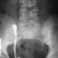
Variable dilation of collecting system to point of obstruction ( Figure 66-1 )
Figure 66-1
Chronic urinary tract obstruction on excretory urography. Grade 3 hydronephrosis and mild parenchymal thinning of the left kidney are seen. The left ureter is dilated down to the level of the sacroiliac joint, where there is irregular narrowing, consistent with a malignant stricture.
(Courtesy WK, Lee, MD, MBBS, St. Vincent’s Hospital, Melbourne, Australia.)
Grading of Hydronephrosis on Excretory Urography
Hydronephrosis can be graded as follows :
- •
Grade 1: Slight blunting of fornices
- •
Grade 2: Obvious blunting of fornices, enlargement of calyces, papillae flattened but visible
- •
Grade 3: Rounded calyces, papillary silhouette obliterated
- •
Grade 4: Extreme ballooning of calyces
EUG has now been largely superseded by ultrasonography, noncontrast computed tomography (CT), and CT and magnetic resonance (MR) urography for assessing UTO. It may still have a limited role in monitoring stone disease.
Retrograde Pyelography
Retrograde pyelography is invasive but demonstrates the collecting system anatomy well. It may be useful when allergy or renal insufficiency preclude the intravenous use of a contrast agent. If the cause of UTO is intraluminal, brushings or biopsy may be performed at the time of retrograde pyelography; ureteral calculi also may be extracted. However, it provides little information about intramural or extraluminal causes of obstruction.
Antegrade Pyelography
Antegrade pyelography is achieved by performing a percutaneous ultrasound-guided renal puncture, followed by injection of iodinated contrast. It is performed before nephrostomy and/or antegrade stenting or, uncommonly, when other imaging methods have failed to demonstrate the cause or site of obstruction.
Digital Subtraction Angiography
Digital subtraction angiography has largely been superseded by multidetector CT (MDCT) angiography. Before the use of CT angiography, digital subtraction angiography was used to determine the course of vessels, especially before operative management of ureteropelvic junction obstruction.
Computed Tomography
Noncontrast Computed Tomography
Noncontrast helical CT was first demonstrated to be equal to EUG in demonstrating ureteral obstruction and more sensitive in diagnosing ureteral calculi by Smith and colleagues in 1995, and it has become the imaging modality of choice for evaluating patients with suspected ureteral colic. It has sensitivity of 95% to 97% and specificity of 96% to 98% for ureteral calculi ( Figure 66-2 ).


