KEY FACTS
Terminology
- •
Urinary tract stone, urinary calculous disease, nephrolithiasis, ureterolithiasis, vesicolithiasis
- •
Macroscopic concretions of crystals in urinary system, sometimes mixed with proteins
Imaging
- •
US has 96% sensitivity, nearly 100% specificity for renal stones > 5 mm
- •
US is valuable for follow-up imaging, particularly in patients with renal colic & known renal stones or patients not improving on treatment for known stone
- •
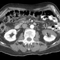
Virtually all stones are visible (including those radiolucent on KUB) on CT except pure matrix stones & protease inhibitor stones (e.g., indinavir, treatment of HIV)
- •
NECT is preferred imaging modality to confirm stone in adult patients with acute flank pain
- ○
Most accurate measurement technique on CT: Bone windows & magnification
- ○
- •
Choice of imaging modality follow-up is often based on visibility of stone on CT scout image
- •
In patients with BMI < 30, consider low-dose CT
- •
Dual-energy CT can differentiate urate stones from other types of stones with high accuracy
Top Differential Diagnoses
- •
Milk of calcium in small cyst
- •
Arterial calcifications
- •
Nephrocalcinosis
- •
Emphysematous pyelonephritis
Pathology
- •
Dependent on type of stone
Clinical Issues
- •
Size, number, location, evidence of obstruction or infection, & relevant anatomic findings (aberrant vasculature, distorted pelvicalyceal architecture, infundibular orientation) are all imaging findings that impact treatment
Scanning Tips
- •
US protocol: Drink 16-32 oz water prior to examination to fill bladder
- •
Always include bladder with special attention to UVJ
- •
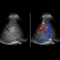
Twinkling artifact on color Doppler is useful to identify otherwise occult stone; more sensitive than acoustic shadowing but higher false-positive rate
 within the mid right kidney. Posterior acoustic shadowing
  helps differentiate this stone from the surrounding sinus fat.
 with twinkling artifact
  and posterior acoustic shadowing
  .
.
Stay updated, free articles. Join our Telegram channel

Full access? Get Clinical Tree


