CHAPTER 39 Vascular Rings and Slings
Vascular rings and slings refer to a spectrum of arterial anomalies caused by abnormalities in development of the embryonic aortic arches.1–4 Complications arise from compression of the trachea, esophagus, or both, leading to respiratory distress and dysphagia. The vast majority of rings and slings are found in infants and young children, but the anomalies can be seen in adults. Computed tomography (CT) and magnetic resonance imaging (MRI) provide accurate anatomic delineation of the vascular anomalies and compressed structures and are the imaging studies of choice to establish the diagnosis. Treatment is surgical intervention for symptomatic patients.
Prevalence and Epidemiology
Vascular rings and slings represent approximately 1% of congenital cardiovascular anomalies,3 although this incidence may be underestimated because some lesions are asymptomatic. Most cases are sporadic, but there may be a genetic inheritance in some arch anomalies. Microdeletions of chromosome 22q11, in particular, have been associated with various arch anomalies.5
Etiology and Pathophysiology
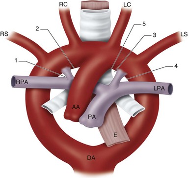
Persistence of a segment of arch that should have regressed or regression of a segment that should have normally persisted explains the development of most arch anomalies. The theoretic embryonic double aortic arch model proposed by Edwards is most extensively used to demonstrate embryologic explanations for the variations in arch development.6 This model classifies vascular rings by the side of the ductus arteriosus and the site of dissolution of the double arches (Fig. 39-1).
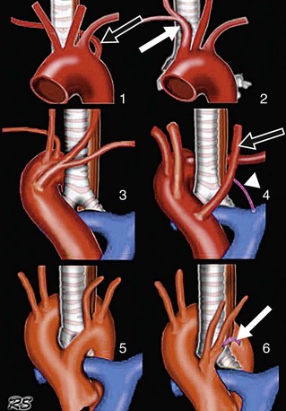
Vascular rings may be complete (true) or incomplete (Fig. 39-2).3 In complete rings, the vascular structures entirely surround and compress the trachea and esophagus. These include the double aortic arch and right arch with aberrant retroesophageal left subclavian artery. In the double arch, an atretic segment of arch may complete the ring. With an aberrant left subclavian, a left ligamentum arteriosum connects the descending aorta and left pulmonary artery completing the ring.
In incomplete rings, the vascular anomalies do not entirely encircle the trachea and esophagus, but do cause mass effect on these structures. These include the left arch with aberrant right subclavian artery and anomalous innominate artery.3 In the case of an aberrant right subclavian, the vessel itself or an aortic diverticulum (Kommerell diverticulum) at the takeoff of the artery compresses the posterior aspect of the esophagus. In anomalous innominate artery, the right innominate artery arises too far to the left from the arch and compresses the trachea anteriorly as it crosses the midline.
MANIFESTATIONS
Clinical Presentation
Symptoms vary with the tightness of the ring around the trachea and/or esophagus, and hence the degree of tracheobronchial compression. Tight rings usually manifest in neonates or infants. Symptoms include stridor, cough, repeated pulmonary infections, cyanosis, and respiratory failure, and feeding difficulties.1–4 Looser rings may be discovered in older children or adults in whom mild dysphagia or choking on food prompts evaluation. Some asymptomatic rings will be discovered incidentally during an imaging study performed for other clinical indications. Rings that are asymptomatic early in life can become symptomatic later in life if the vascular structures become ectatic and compress the airway or esophagus.2 Double aortic arches tend have more severe symptoms and present earlier than other rings.7 Slings commonly manifest in neonates, producing respiratory compromise.
Imaging Studies
Techniques and Findings
Radiography
Chest radiography is used to show the side of the aortic arch and compression of adjacent structures. If a right arch is identified, the likelihood of a vascular ring is high. If only a left arch is identified, a vascular ring is less likely, but not excluded.2,8
Computed Tomography
Computed tomography can provide accurate anatomic detail of the vascular anomalies and compressed trachea or esophagus. CT is especially useful for the evaluation of the tracheobronchial tree, simulating the surgical view.1
Stay updated, free articles. Join our Telegram channel

Full access? Get Clinical Tree


 FIGURE 39-1
 FIGURE 39-2