Chapter 188
Venous Malformations
Epidemiology
Venous malformations are categorized as a type of vascular malformation using the classification system of Mulliken and Glowacki. Most authors now classify what were once termed cavernous hemangiomas as venous malformations. These lesions are felt to be present at birth but are usually detected in late childhood or early adulthood.
Clinical Findings
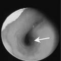
Small lesions may present as an asymptomatic bluish mass that is easily compressible. More advanced lesions may be painful, may result in functional impairment, or hemorrhage. The lesions may enlarge with patient position or with crying. The lesions may also enlarge with Valsalva’s maneuver or following a placement of a tourniquet to obstruct the venous out-flow. The most common locations in the extracranial head and neck include subcutaneous tissues of the face, muscles of mastication, periorbital region and deep neck spaces.
Pathology
These lesions arise from anomalous venous development. The absence of valves result in stagnant flow and variable communication with the surrounding normal venous system. Enlargement of the lesion over time is due to growth of the patient rather than to endothelial proliferation.
Treatment
Complete surgical resection is the treatment of choice for localized lesions with well-defined margins. Because these are low flow venous lesions, percutaneous sclerotherapy is becoming an accepted treatment option for patients with advanced infiltrative lesions.
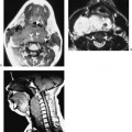
Imaging Findings
US