Fig. 1
Non-contrast-enhanced CT of the brain at presentation (a, b) and after clinical deterioration on the day of the angiography (c, d). (a) This non-contrast CT shows hyperdensity of the vein of Galen and the straight sinus consistent with thrombotic occlusion. Not the normal density of the superior sagittal sinus. There is no abnormality of the brain parenchyma, especially not of the thalami. (b) This CT at a more caudal location shows a hyperdense left sigmoid sinus and a normal right sigmoid. (c) This non-enhanced CT scan before and after the patient became unconscious shows prominent thalamic edema bilaterally. The low-density changes extend into the medial basal ganglia on the left. There is high density of the internal cerebral veins, the vein of Galen, and the straight sinus. (d) This slice shows now a large hyperdense thrombus in the left transverse sinus. As this clot propagation had occurred under intravenous heparin medication, the patient was referred for endovascular treatment with selective thrombolysis and clot aspiration
CVT was diagnosed, and low-molecular-weight heparin was started, but she continued to deteriorate to unconsciousness. Repeat CT study the next day showed progression of the sinus thrombus to involve the internal cerebral veins with bilateral thalamic edema (Fig. 2).
Get Clinical Tree app for offline access
Fig. 2
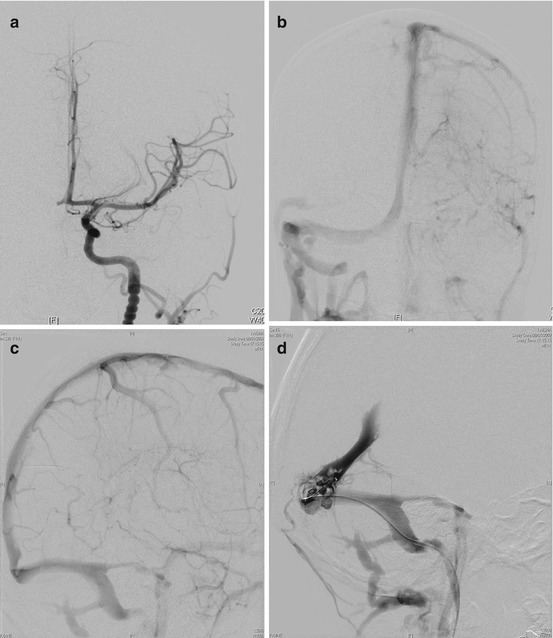
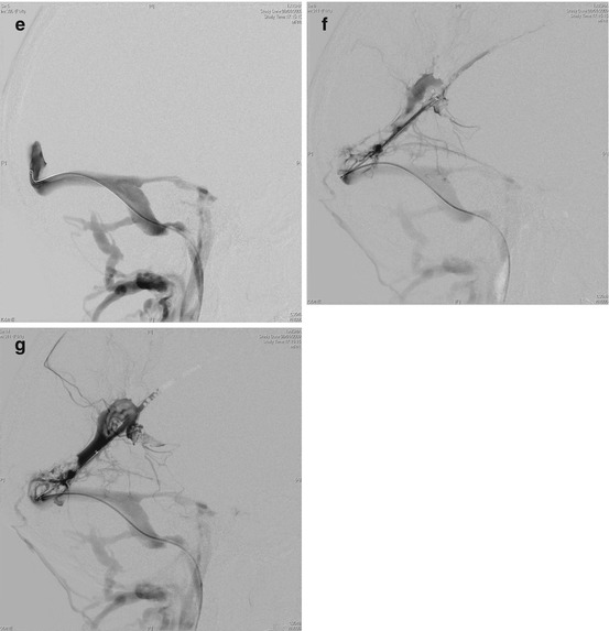
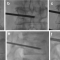
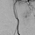
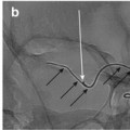
Cerebral angiogram under general anesthesia. Right venous approach (6 F sheath in the femoral vein) and left arterial approach (5 F sheath in the femoral artery). (a) Left internal carotid artery injection, arterial phase, and anterior posterior projection. There is a conspicuous “string of pearls” appearance of the distal left ICA suggestive of homocystinuria. (b) The venous phase of the same injection does not show the left transverse and sigmoid sinus, which are occluded by thrombus. (c) The lateral view of the left ICA injection fails to demonstrate the straight sinus and the internal cerebral veins. Only one transverse and sigmoid sinus is visible. (d) A Vasco 35 microcatheter is seen in the confluens sinuum adjacent to a clot protruding from the straight sinus into the confluens. (e


Related posts:
Stay updated, free articles. Join our Telegram channel
Full access? Get Clinical Tree
