Viruses are the most common cause of respiratory infection and may result in rhinitis, pharyngitis, laryngotracheitis, bronchitis, bronchiolitis, and, less commonly, pneumonia. Most viral pneumonias in immunocompetent adults are due to influenza viruses; other common viral etiologies include respiratory syncytial virus (RSV) and adenovirus. Immunocompromised hosts are particularly susceptible to pneumonias caused by cytomegalovirus (CMV) and herpesviruses. Risk factors associated with increased incidence and severity of viral infections include very young and old age, crowding, malnutrition, and immunologic disorders. Respiratory viral infections are a much greater problem in developing countries than in developed countries and are one of the leading causes of death in children younger than 5 years. In addition to these acute complications, viral-induced childhood bronchiolitis is well recognized as a precursor of adult bronchiectasis and constrictive bronchiolitis. Airway infection can also induce symptoms of asthma in patients who have this disease, and there is evidence that such infection in childhood is involved in the pathogenesis of asthma in later life. Finally, several viruses, such as Epstein-Barr virus (EBV), herpesvirus 8, and papillomavirus, are implicated in the development of some pulmonary and pleural neoplasms.
The diagnosis of viral pneumonia is often one of exclusion and is based on an absence of sputum production, a failure to culture pathogenic bacteria, a relatively benign clinical presentation, a white blood cell count or procalcitonin level that is normal or only slightly elevated, a chest radiograph that reveals bronchopneumonia or localized interstitial disease, and a lack of response to antibiotic therapy. Confirmation of the diagnosis and identification of the specific causative virus can be accomplished by a variety of means, including culture, serology, detection of viral antigens within respiratory tract secretions or blood by monoclonal antibodies, detection of virus-associated molecules by in situ hybridization or polymerase chain reaction (PCR), and observation of virus-induced changes cytologically or histologically. The sensitivity and specificity of these tests vary and are related to some extent to the underlying organism.
Patterns of Disease
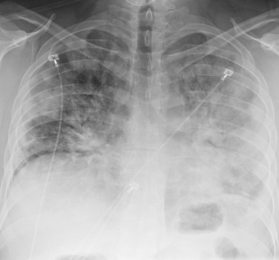
The radiographic manifestations of viral infection are protean and cannot be reliably differentiated from other infections caused by bacterial organisms. Chest radiographs may be normal or often show unilateral or bilateral foci of consolidation, ground-glass opacities, nodular opacities, and bronchial wall thickening ( Figs. 13.1 and 13.2 ). Lobar consolidation, lymphadenopathy, and pleural effusions are uncommon and are not seen in most cases of viral pneumonia.


A retrospective study compared the computed tomography (CT) appearances of 93 viral pneumonias and 22 bacterial pneumonias in both immunocompetent and immunocompromised hosts to determine CT patterns that may allow a specific diagnosis. Viral or bacterial pneumonia was determined to be present if a patient had lower respiratory tract symptoms and laboratory evidence of infection. Viral pneumonias manifested with one of five different CT patterns:
- 1.
A normal CT or a CT with abnormalities unrelated to infection (e.g., linear scar, emphysema, etc.) was the most common CT appearance observed in 35% of patients with viral pneumonia.
- 2.
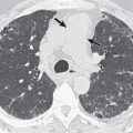
Airway centric disease was the second most common CT appearance observed in 33% of patients ( Fig. 13.3 ). Typical features include bronchial wall thickening, bronchiolitis with centrilobular nodules or tree-in-bud opacities, and peribronchial consolidation or ground-glass opacities. Centrilobular nodules typically measure less than 10 mm. Other frequent CT abnormalities that often coexist with airway centric disease include mosaic attenuation, lung hyperinflation, and expiratory air trapping.

Fig. 13.3
Airway centric pattern with parainfluenza pneumonia. Axial CT shows bilateral bronchial wall thickening (arrowheads), patchy ground-glass opacities, and tree-in-bud opacities (arrow) . Note focal areas of decreased lung attenuation (asterisks) representing mosaic attenuation caused by small airways infection and secondary air trapping.
(From Walker CM, Digumarthy S. Pulmonary infections. In: Digumarthy S, Abbara S, Chung JH, eds. Problem Solving in Chest Imaging. Philadelphia: Elsevier; 2019.)
- 3.
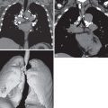
Multifocal airspace pattern was the third most common CT appearance observed in 24% of patients ( Fig. 13.4 ). Typical features include multifocal ground-glass opacities and/or consolidation with intervening normal portions of lung. Of importance, bronchial abnormalities, including tree-in-bud opacities, are generally minimal or absent.

Fig. 13.4
Multifocal airspace pattern with adenovirus pneumonia. Axial CT shows multifocal consolidation and ground-glass opacities.
- 4.
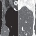
Unifocal airspace pattern was uncommon, affecting 6% of patients ( Fig. 13.5 ). Typical features include focal consolidation, ground-glass opacities, or tree-in-bud opacities.

Fig. 13.5
Unifocal airspace pattern with influenza pneumonia. Axial CT at the level of the right upper lobe bronchus shows focal peribronchial consolidation and small nodular opacities.
- 5.
Diffuse airspace pattern was rare, affecting 2% of patients ( Fig. 13.6 ). Typical features include diffuse, relatively uniform bilateral ground-glass opacities, or consolidation.

Fig. 13.6
Diffuse airspace pattern with influenza A pneumonia. Axial CT shows bilateral ground-glass opacities with peribronchial consolidation in the right lower lobe. The findings affected both lungs diffusely.
When these patterns were compared with patients with bacterial pneumonia, the diffuse disease pattern and the presence of a pleural effusion were more commonly associated with bacterial pneumonia, and only the normal CT pattern was more commonly associated with viral pneumonia. Otherwise, the imaging appearances overlapped considerably, and other methods, including patient presentation and laboratory analysis, were used in conjunction with the imaging findings to make a specific diagnosis.
Interlobular septal thickening ( Fig. 13.7 ) is an uncommon manifestation that is usually seen only in certain infections, such as hantavirus or in viral infection that progresses to acute respiratory distress syndrome (ARDS). Significant lymphadenopathy and moderate or large pleural effusions are uncommon in viral infection and should prompt search for an alternative etiology.

Specific Viruses
Influenza Viruses
Etiology, Prevalence, and Epidemiology
Influenza viruses are single-stranded ribonucleic acid (RNA) viruses found worldwide. They are subdivided into three antigenic types A, B, and C. Influenza can occur in pandemics, epidemics, or sporadically in individuals or small clusters of patients. Type A viruses cause almost all severe epidemics and all pandemics. These organisms have the ability to change their structural and antigenic nature, the variant forms possessing different virulence from that of their progenitors.
Although the type A virus is generally transmitted from person to person by droplet infection, antigenically similar viruses also infect swine, horses, and wild and domesticated birds, and human disease is occasionally derived from these sources. It is believed that these animals serve as a milieu for genetic recombination whereby new and sometimes more virulent strains capable of causing human disease are created. Influenza is an important infection worldwide, being estimated to result in symptomatic disease in approximately 20% of children and 5% of adults each year.
Influenza outbreaks tend to occur on an annual basis, typically during the winter in temperate climates; in tropical and subtropical areas, they occur either during the rainy season or throughout the year, depending on the particular location. Attack rates are especially high in schoolchildren; complications and hospitalization also are more likely in these individuals and in the elderly. The 24- to 48-hour incubation period allows rapid spread of the disease. It is highly contagious, and a large proportion of individuals may contract the disease to some degree in closed environments, such as on cruise ships and airplanes. Nosocomial transmission of disease is well documented.
Pneumonia is an uncommon but serious complication of influenza infection that is usually caused by type A and occasionally type B organisms. Although it is often mild, it can be overwhelming and rapidly fatal, with most cases recognized during epidemics or pandemics. In approximately one-third of cases of severe pneumonia, the illness develops abruptly in apparently healthy individuals. Most of the remainder have a predisposing condition, such as cardiac disease, pregnancy, cystic fibrosis, or immunodeficiency. Infants and the elderly are at particular risk for serious disease. Death can result from severe influenza pneumonia or from superinfection by bacteria, such as Staphylococcus aureus, Streptococcus pneumoniae, Haemophilus influenzae, and Moraxella catarrhalis.
Clinical Presentation
The most common manifestations of influenza infection consist of rapid onset of dry cough, myalgia, chills, headache, conjunctivitis, and fever of greater than 38°C. In older patients and patients with underlying cardiopulmonary disease, there is a tendency for involvement of the lower respiratory tract, with development of bronchitis, bronchiolitis, and pneumonia. Patients who have pneumonia tend to have more severe cough and may develop severe dyspnea, cyanosis, and hypoxemia.
Bacterial superinfection usually develops within a few days after the initial viral infection. Superimposed bacterial infection occurs most commonly in the elderly and in patients with underlying pulmonary disease. Clinically, these patients initially present with typical influenza symptoms. The symptoms seem to be improving when the clinical course is complicated by recrudescence of fever and development of chills, pleuritic chest pain, and productive cough. The symptoms in these patients tend to be milder than in patients with primary influenza pneumonia, and the mortality is lower.
Pathophysiology
Transmission of influenza viruses is by airborne droplets and may result in rhinitis, pharyngitis, laryngitis, tracheobronchitis, bronchiolitis, and bronchopneumonia. The histologic appearance of fatal influenza pneumonia is diffuse alveolar damage, the parenchyma showing a variably severe interstitial mononuclear inflammatory infiltrate; consolidation of airspaces by edema, hemorrhage, and fibrin; and hyaline membranes. Less severe cases of pneumonia may result in findings of mild acute lung injury and organizing pneumonia. The virus can be cultured or seen by immunofluorescence in a large proportion of cases.
Manifestations of the Disease
Radiography
The most common radiographic manifestations of influenza pneumonia consist of bilateral reticulonodular opacities with or without superimposed areas of consolidation ( Fig. 13.8 ). Less commonly, patients with primary influenza pneumonia may present with focal areas of consolidation, usually in the lower lobes, without apparent reticular or reticulonodular opacities. Serial radiographs may show poorly defined, patchy or nodular areas of consolidation, 1 to 2 cm in diameter, which become rapidly confluent ( Fig. 13.9 ). Pleural effusion is uncommon. The radiologic abnormalities usually resolve in approximately 3 weeks. The manifestations of secondary bacterial pneumonia are the same as bronchopneumonia and consist of lobular, subsegmental, or segmental unilateral or bilateral areas of consolidation.


Computed Tomography
Miller and associates described the CT findings of influenza infection in 60 patients. Influenza had a myriad of imaging appearances comprising a normal CT (43%), an airway centric pattern manifesting with bronchial wall thickening, centrilobular nodules, and tree-in-bud opacities (27%), a multifocal airspace pattern with multifocal consolidation and/or ground-glass opacities (20%) (see Fig. 13.8 ), a focal airspace pattern with focal ground-glass opacity or consolidation (8%), and a diffuse airspace pattern with bilateral ground-glass opacities or consolidation (2%) ( Fig. 13.10 ). The centrilobular nodules seen with influenza often measure 2 to 9 mm in diameter and are usually bilateral and asymmetric in distribution (see Fig. 13.8 ).

Avian influenza strain H5N1, swine influenza strain H1N1, and most recently a new avian influenza strain H7N9 have emerged with periodic outbreaks related to cross-species transmission after humans acquire the virus after being in close contact with infected birds or pigs or their products. Infection by these influenza viruses preferentially affects younger, previously healthy individuals, and transmission of the H1N1 strain was declared to be in a worldwide pandemic state by the World Health Organization in 2009. The case fatality rate is usually high and has approached 60% in patients infected by certain strains. The CT appearance of avian influenza consists of focal, multifocal, or diffuse ground-glass opacities or consolidation. Pleural effusions, lymphadenopathy, centrilobular nodules, pseudocavitation, and pneumatocele formation are often also noted. Swine influenza (H1N1) often resembles organizing pneumonia, manifesting as peribronchial or peripheral predominant consolidation and ground-glass opacities ( Fig. 13.11 ). Of importance, patients with H1N1 infection also have an elevated risk of pulmonary thromboembolic disease.
- •
Symptomatic disease (influenza) occurs in 20% of children and 5% of adults per year.
- •
Risk factors for primary influenza virus pneumonia include old age and cardiopulmonary disease.
- •
The most common imaging manifestations of primary influenza virus pneumonia follow:
- •
Reticulonodular pattern
- •
Focal areas of consolidation that may become confluent
- •
Peripheral or peribronchial consolidation that resembles organizing pneumonia in some infected with H1N1 strain
- •
- •
Secondary bacterial pneumonia is relatively common and most commonly affects the elderly and those with underlying cardiopulmonary disease.
- •
The most common radiologic manifestation of secondary bacterial pneumonia is patchy or confluent unilateral or bilateral consolidation.

Respiratory Syncytial Virus and Parainfluenza Virus
Etiology, Prevalence, and Epidemiology
Respiratory syncytial virus (RSV) and parainfluenza virus account for a high proportion of cases of pneumonia, acute bronchiolitis, and tracheobronchitis (croup) in young children ( Fig. 13.12 ) and occasionally cause lower respiratory tract infection in adults. Both are single-stranded RNA viruses and members of the Paramyxoviridae family.

RSV infects virtually all children in the first few years of life and is the most common cause of bronchiolitis and one of the most common causes of pneumonia in these patients. It has been estimated to be responsible for about 90,000 hospitalizations and 4500 deaths of infants and young children per year in the United States. Both viruses are being recognized increasingly in adults, particularly adults who have underlying cardiopulmonary disease, malignancy, or immunodeficiency or who are institutionalized. RSV is highly contagious and may result in high infection rates in relatively enclosed places, such as day care centers and nursing homes.
Clinical Presentation
Symptoms of infection in adults are usually those of a “cold” (rhinitis, pharyngitis, and conjunctivitis). Manifestations of lower respiratory tract involvement (most often cough) are seen in a significant number of patients.
Pathophysiology
Transmission is by airborne droplets or hand-to-surface contact. Viral infection may involve mainly the airways or less commonly the parenchyma. When airway involvement predominates, the most severe changes occur in small membranous and respiratory bronchioles and consist of epithelial degeneration accompanied by a variably intense inflammatory cell infiltrate. Parenchymal involvement results in interstitial pneumonitis or diffuse alveolar damage.
Manifestations of the Disease
Radiography
The radiographic findings in infants and children consist of bronchial wall thickening and peribronchial (central) areas of consolidation (see Fig. 13.1 ). Other common findings include hyperinflation (reflecting the presence of acute bronchiolitis) and patchy bilateral consolidation (reflecting the presence of bronchopneumonia).
The radiographic findings of RSV pneumonia in adults usually consist of patchy bilateral areas of consolidation. Less commonly, patients may present with a bilateral reticulonodular pattern. Rarely, patients may develop acute pneumonia with rapid progression to ARDS.
Computed Tomography
The CT findings of RSV and parainfluenza virus are similar, usually manifesting with an airway centric pattern of disease consisting of bronchial wall thickening, centrilobular nodules, and branching nodular opacities (tree-in-bud pattern), reflecting the presence of bronchiolitis (see Fig. 13.3 ). There are often peribronchiolar ground-glass opacities or areas of consolidation, owing to bronchopneumonia ( Fig. 13.13 ). Inspiratory CT may show mosaic attenuation, and expiratory CT may show air trapping. Less common manifestations include multifocal airspace or focal airspace patterns without appreciable bronchial or small airways involvement.
- •
Infection with RSV and parainfluenza virus is common in infants and children; infection is less common in adults.
- •
Risk factors for RSV bronchiolitis include young age (infants and young children).
- •
The most common radiologic manifestations include:
- •
Hyperinflation
- •
Bronchial wall thickening and peribronchial opacities
- •
Steeple sign in croup
- •
- •
Risk factors for pneumonia include the extremes of age (infants, young children, the elderly) and chronic illness.
- •
The most common CT manifestations include:
- •
Airway centric pattern of disease with bronchial wall thickening, centrilobular nodules, and tree-in-bud opacities
- •
Patchy peribronchial consolidation or ground-glass opacities
- •

Severe Acute Respiratory Syndrome Coronavirus
Etiology, Prevalence, and Epidemiology
The SARS coronavirus is the cause of severe acute respiratory syndrome (SARS), an infectious disease that first emerged in southern China in 2002. When it reached Hong Kong, the disease spread rapidly to other parts of the world by international air travel. Within approximately 8 months, SARS affected 8422 patients in numerous countries and resulted in 916 deaths (case-fatality rate of 11%). Most of the infections occurred in hospitals, laboratories involved in diagnosis or research on the organism, and nursing homes. Only a few sporadic cases of SARS have been reported since 2003.
The natural reservoir of the organism is believed to be wild animals, such as raccoon-dogs, ferrets, and civets. The disease is transmitted by droplets or direct inoculation from contact with infected surfaces. The mean incubation period is 6 days (range, 2–10 days).
Clinical Presentation
Presenting symptoms include fever, chills, dry cough, myalgia, and headache. These can progress to clinical, radiologic, and pathologic features of ARDS. Laboratory findings include lymphopenia, evidence of disseminated intravascular coagulation, and elevated blood levels of lactate dehydrogenase and creatine kinase.
Pathophysiology
The predominant pattern of lung injury in autopsy specimens was diffuse alveolar damage. Cases of 10 or fewer days of duration showed acute-phase diffuse alveolar damage. Cases of more than 10 days of duration exhibited organizing-phase diffuse alveolar damage and, frequently, superimposed bacterial bronchopneumonia.
Manifestations of the Disease
Radiography
The most common radiologic manifestations consist of focal unilateral or multifocal unilateral or bilateral areas of consolidation ( Fig. 13.14 ). The consolidation tends to involve predominantly the peripheral lung regions and the middle and lower lung zones. Less common radiographic findings include focal or diffuse ground-glass opacities and, rarely, lobar consolidation. Approximately 20% to 40% of patients with SARS have normal radiographs at presentation.


