, Lawrence A. Zumo2 and Valerie Sim3
(1)
Parkinson’s Clinic of Eastern Toronto, Toronto, ON, Canada
(2)
Silver Spring, Cheverly, MD, USA
(3)
Centre for Prions and Protein Folding Diseases, University of Alberta, Edmonton, AB, Canada
Abstract
There are many diseases and syndromes which affect the white matter of the brain. The most common by far is multiple sclerosis, but the differential includes infectious, inflammatory, metabolic, mitochondrial, vascular, neoplastic and toxic etiologies. In this chapter we present the 2010 McDonald MRI diagnostic criteria for multiple sclerosis and include example cases of mulitple sclerosis, transverse myelitis, autoimmune encephalitis, toxic demyelination, posterior reversible encephalopathy syndrome and neuroBechet’s disease. Progressive multifocal leukoencephalopathy is presented under infectious diseases (Chap. 9), CNS lymphoma is presented under Tumours (Chap. 4), and osmotic demyelination is presented under metabolic disorders (Chap. 10).
McDonald Criteria for Diagnosis of Multiple Sclerosis
For a patient to be diagnosed with multiple sclerosis, he or she must have evidence of demyelination which is disseminated in space and time. MRI can support the diagnosis, as defined the McDonald criteria, most recently revised in 2010 (Polman et al. 2011). Separation in space is met by one or more hyperintense lesions on T2-weighted imaging in at least two of four regions (periventricular, juxtacortical, infratentorial or spinal cord). Separation in time is met by the simultaneous presence of asymptomatic enhancing and non-enhancing lesions, or new T2 or enhancing lesions on subsequent MRI scans.
Case 3.1 Multiple Sclerosis (FLAIR Sequences)
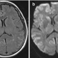
A 43 year old right-handed female presented with progressive numbness from her right axilla to her trunk to both legs. There was no problem with her vision, speech, or swallowing. She had no weakness, bowel or bladder symptoms. Her symptoms resolved over the next 4 weeks. Three months later she developed persistent numbness on the left side, which resolved after 3 weeks. An MRI was performed (Fig. 3.1).
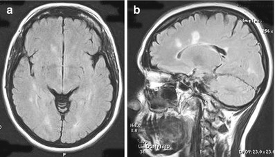

Figure 3.1
Axial (a) and sagittal (b) FLAIR MRI showing multiple areas of demyelination
Explanation and Diagnosis
Figure 3.1 shows multiple asymmetrical oval hyperintense signal abnormalities which are disseminated in space and are consistent with demyelination. Their ovoid appearance and projection from the corpus callosum (known as Dawson fingers) are typical for multiple sclerosis.
Case 3.2 Multiple Sclerosis (T1 and T2 Sequences)
A 27 year old right-handed male presented with a gradual onset of blurred vision and numbness on the right side of his body which lasted for 3 weeks, after which the numbness resolved gradually. An MRI was performed (Fig. 3.2).
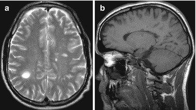

Figure 3.2
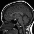
(a) Axial T2-weighted MRI showing multiple areas of demyelination; (b) sagittal T1-weighted MRI showing multiple hypointense signal abnormalities (black holes) involving the corpus callosum and subcortical white matter
Explanation and Diagnosis
< div class='tao-gold-member'>
Only gold members can continue reading. Log In or Register to continue
Stay updated, free articles. Join our Telegram channel

Full access? Get Clinical Tree


