KEY FACTS
Terminology
- •
Rare inflammatory process causing focal or diffuse destruction of gallbladder (GB) wall with accumulation of lipid-laden macrophages, fibrous tissue, and acute and chronic inflammatory cells
Imaging
- •
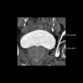
Marked GB wall thickening
- ○
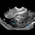
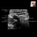
Intramural hypoechoic nodules or bands with continuous mucosa
- –
Nodular areas of foamy inflammatory cells or necrosis/abscess
- –
- ○
- •
Gallstones in 80%
- •
Absence of hepatic invasion or biliary dilatation when uncomplicated
- •
Complications in 30%
- ○
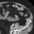
Infiltrative form: GB fossa mass involving adjacent organs and surrounding fat/soft tissue obliterating normal margins, preoperative differentiation from carcinoma nearly impossible
- ○
Abscesses, fistula
- ○
Top Differential Diagnoses
- •
GB carcinoma
- •
Gangrenous cholecystitis
- •
Hyperplastic cholecystoses
Clinical Issues
- •
1-2% of cholecystectomy specimens
- ○
Adenocarcinoma seen in up to 10% of resected specimens
- ○
- •
Mean age at presentation: 44-63 years; F > M
- •
Symptoms of chronic cholecystitis most common, followed by symptoms of acute cholecystitis with leucocytosis
- •
Less common presentation: Obstructive jaundice, cholangitis, palpable mass
- •
Treatment: Open cholecystectomy
Scanning Tips
- •
Look for intramural hypoechoic nodules and preservation of mucosal line, which favor this over carcinoma