KEY FACTS
Terminology
- •
Chronic renal inflammation usually associated with longstanding urinary calculus obstruction (75%)
- •
Renal parenchyma is gradually replaced by lipid-laden macrophages
- •
Diffuse (> 80%) and focal (< 20%) forms
- •
3 stages of xanthogranulomatous pyelonephritis: Intrarenal → perirenal → perinephric ± retroperitoneal involvement
Imaging
- •
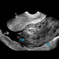
Diffusely enlarged kidney with hypoechoic round masses replacing normal parenchyma on US
- •
“Staghorn” calculus with renal enlargement and perirenal fibrofatty proliferation
- •
Renal sinus fat obliterated with large central “staghorn” calculus
- •
Perinephric extension ± adjacent organs or structures may include sinus tracts or abscesses
- •
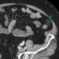
Multiple focal, low-attenuating renal masses with rim enhancement on CT
- •
Ultrasound ideal at initial investigation; CT good for assessing excretory function and retroperitoneal involvement
Pathology
- •
Lipid-laden, “foamy” macrophages, diffuse infiltration of plasma cells and histiocytes
Clinical Issues
- •
Flank pain, fever, palpable mass, and weight loss
- •
Rare complications: Hepatic dysfunction, extrarenal extension, fistulas
- •
Long-term chronic process with good prognosis if treated and rare mortality
- •
Antibiotic treatment is sometimes effective
- •
Severe disease or perinephric extension usually requires nephrectomy
 , causing replacement of parenchyma by collections of foamy macrophages
  .
.