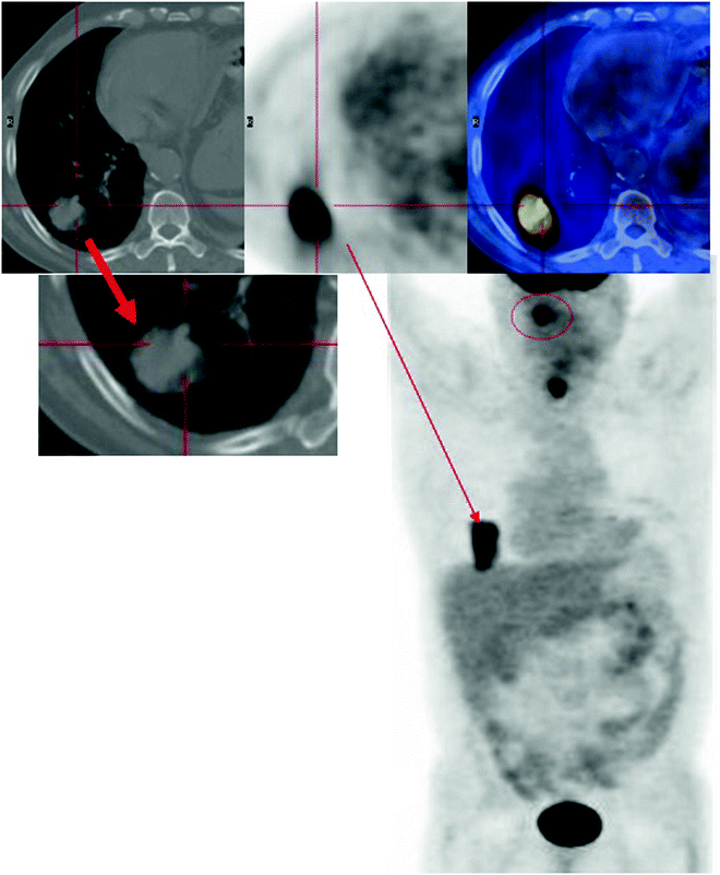
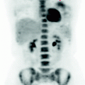
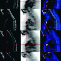
Fig. 8.1
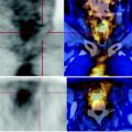
Diffuse thickening of the laryngeal mucosa, with no solid focal nodular lesions seen
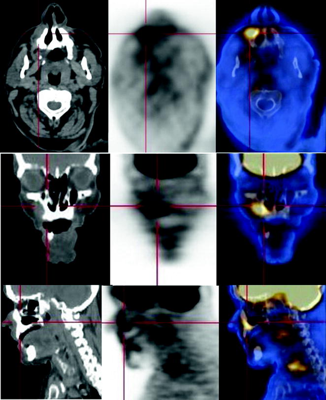
Fig. 8.2
CT scans show a diffuse thickening of the laryngeal mucosa, with no solid focal nodular lesions seen
The PET scan shows widespread and increased concentration of FDG that does not resemble the typical features of nodular malignant processes.
8.4 Conclusions
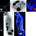
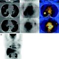
The PET scan shows the right pulmonary neoplasm with a high metabolism and a skin lesion in the ipsilateral zygomatic region, which is also metabolically active, consistent with a diagnosis of synchronous squamous cell carcinoma (Fig. 8.3).
