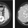
CASE 15 A 45-year-old Caucasian woman presents with recurrent right upper quadrant pain made worse after having fatty food. Fig. 15.1 Axial contrast-enhanced CT image shows an annular area of narrowing in the gallbladder with dumbbell-shaped compartmentalization. There are associated outpouchings in the gallbladder wall and thickening. An axial contrast-enhanced computed tomography (CT) image shows an annular area of narrowing in the gallbladder with dumbbell-shaped compartmentalization. There are associated outpouchings in the gallbladder wall and thickening (Fig. 15.1). Adenomyomatosis of gallbladder Adenomyomatosis is found in 3% of the cholecystectomy specimens and is associated with thickening of the gallbladder wall with the presence of intramural diverticuli, which may contain bile, biliary sludge, or cholesterol crystals. Adenomyomatosis may be associated with cholesterolosis in some cases.
Clinical Presentation

Radiologic Findings
Diagnosis
Differential Diagnosis
Discussion
Background
Clinical Findings
Related posts:


Stay updated, free articles. Join our Telegram channel

Full access? Get Clinical Tree


