CASE 43 A 68-year-old woman presents with a 1-week history of exertional chest pain and shortness of breath. She is found to have heart failure, with an ejection fraction of 20%. The troponin level is elevated to 0.16 ng/mL, with lateral T-wave inversions on ECG. She is referred for PET to assess for myocardial viability and potential coronary revascularization. Fig. 43.1 • The patient had nothing to eat within 4 hours of the test. • After a scout CT acquisition (120 kVp, 10 mA) for patient positioning, a CT transmission scan (140 kVp, 30 mA, pitch of 1.35) was acquired for attenuation correction. Commercial software was used for coregistration of the transmission and emission images. • The patient underwent a rest myocardial perfusion PET study. Rest emission images were obtained after the intravenous administration of 60 mCi of 82Rb, with imaging starting at 120 seconds (given the low ejection fraction) after completion of the radionuclide infusion and continued for 5 minutes. Rest gated images were acquired at 8 frames per cycle. • PET images were reconstructed with ordered subsets expectation maximization (OSEM; 2 iterations and 30 subsets), and a three-dimensional PET filter was used (Butterworth filter cutoff frequency of 10, order of 5). • After the rest image acquisition, the patient received 25 g of oral Trutol (glucose tolerance beverage) and a total of 9 U of regular insulin intravenously to optimize myocardial utilization of glucose.
Clinical Presentation
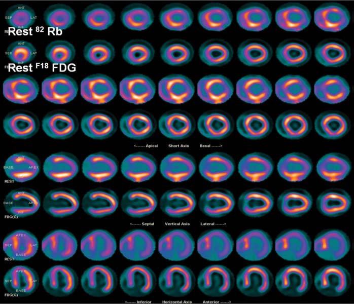
Technique
Related posts:


Stay updated, free articles. Join our Telegram channel

Full access? Get Clinical Tree


